{"title":"Morphological development of the ovary in the <i>Alectoris chukar</i> at embryonic and pre-pubertal stages.","authors":"Morvarid Teymouri, Masoumeh Kheirabadi, Abolghasem Nabipour","doi":"10.30466/vrf.2024.2035219.4357","DOIUrl":null,"url":null,"abstract":"<p><p><i>Alectoris chukar</i> (AC) is a common model organism in biological research. To understand oogenesis and folliculogenesis mechanisms in bird reproduction, we analyzed the ovarian tissue structure of AC at embryonic and pre-pubertal stages. Fertilized eggs, newborn chicks and juvenile AC were used to study the tissue structure of female gonads. Sections of ovaries were prepared and examined using various histological techniques including Hematoxylin and Eosin, Periodic acid-Schiff and Masson's trichrome. Semi-thin and ultra-thin sections of ovary in newly-hatched chicks were prepared for study by electron microscope. The study revealed asymmetry between the left and right ovaries, with a larger left ovary. The functional left ovary exhibited a cortex and medulla, containing somatic and germ cells, with an increase in germ cell number, size and volume leading to cortex thickening. Meiosis division of germ cells and oocyte formation were observed with pre-follicular cells surrounding them. Electron microscopy revealed mitochondria and desmosome cell junctions in germ cells. Our study provided insights into tissue changes in ovaries and germ cells at different developmental stages of AC embryos, newly-hatched chicks and juvenile AC. The results suggested that cortex thickening and germ cell mitochondria density could be used as hallmarks of healthy AC maturity under normal physiological conditions. Further research should explore the impact of growth factors, hormones and environmental factors to unravel avian ovarian development complexities and improve AC reproductive biology knowledge.</p>","PeriodicalId":23989,"journal":{"name":"Veterinary Research Forum","volume":"16 6","pages":"39-344"},"PeriodicalIF":1.1000,"publicationDate":"2025-01-01","publicationTypes":"Journal Article","fieldsOfStudy":null,"isOpenAccess":false,"openAccessPdf":"https://www.ncbi.nlm.nih.gov/pmc/articles/PMC12295533/pdf/","citationCount":"0","resultStr":null,"platform":"Semanticscholar","paperid":null,"PeriodicalName":"Veterinary Research Forum","FirstCategoryId":"97","ListUrlMain":"https://doi.org/10.30466/vrf.2024.2035219.4357","RegionNum":4,"RegionCategory":"农林科学","ArticlePicture":[],"TitleCN":null,"AbstractTextCN":null,"PMCID":null,"EPubDate":"2025/6/15 0:00:00","PubModel":"Epub","JCR":"Q3","JCRName":"ZOOLOGY","Score":null,"Total":0}
引用次数: 0
Abstract
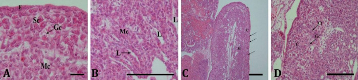
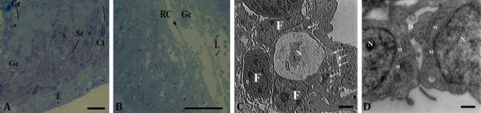
Alectoris chukar (AC) is a common model organism in biological research. To understand oogenesis and folliculogenesis mechanisms in bird reproduction, we analyzed the ovarian tissue structure of AC at embryonic and pre-pubertal stages. Fertilized eggs, newborn chicks and juvenile AC were used to study the tissue structure of female gonads. Sections of ovaries were prepared and examined using various histological techniques including Hematoxylin and Eosin, Periodic acid-Schiff and Masson's trichrome. Semi-thin and ultra-thin sections of ovary in newly-hatched chicks were prepared for study by electron microscope. The study revealed asymmetry between the left and right ovaries, with a larger left ovary. The functional left ovary exhibited a cortex and medulla, containing somatic and germ cells, with an increase in germ cell number, size and volume leading to cortex thickening. Meiosis division of germ cells and oocyte formation were observed with pre-follicular cells surrounding them. Electron microscopy revealed mitochondria and desmosome cell junctions in germ cells. Our study provided insights into tissue changes in ovaries and germ cells at different developmental stages of AC embryos, newly-hatched chicks and juvenile AC. The results suggested that cortex thickening and germ cell mitochondria density could be used as hallmarks of healthy AC maturity under normal physiological conditions. Further research should explore the impact of growth factors, hormones and environmental factors to unravel avian ovarian development complexities and improve AC reproductive biology knowledge.
期刊介绍:
Veterinary Research Forum (VRF) is a quarterly international journal committed to publish worldwide contributions on all aspects of veterinary science and medicine, including anatomy and histology, physiology and pharmacology, anatomic and clinical pathology, parasitology, microbiology, immunology and epidemiology, food hygiene, poultry science, fish and aquaculture, anesthesia and surgery, large and small animal internal medicine, large and small animal reproduction, biotechnology and diagnostic imaging of domestic, companion and farm animals.

 求助内容:
求助内容: 应助结果提醒方式:


