Qinqin Yan, Fuhua Yan, Qi Lin, Qiqi Cao, Yajie Zhang, Xiaoyan Chen, Bernhard Schmidt, Zhihan Xu, Wenjie Yang
{"title":"Low-dose ultrahigh-resolution PCCT enhances subsolid nodule characterization.","authors":"Qinqin Yan, Fuhua Yan, Qi Lin, Qiqi Cao, Yajie Zhang, Xiaoyan Chen, Bernhard Schmidt, Zhihan Xu, Wenjie Yang","doi":"10.1007/s11547-025-02057-0","DOIUrl":null,"url":null,"abstract":"<p><strong>Purpose: </strong>To characterize invasion-associated CT features in pulmonary subsolid nodules using low-dose ultrahigh-resolution (UHR) photon-counting CT (PCCT) images and evaluate UHR's diagnostic superiority over standard high-resolution (HR) images.</p><p><strong>Methods: </strong>Patients with subsolid lung adenocarcinoma were recruited for chest scan on PCCT to obtain UHR and standard HR images between November 2023 and May 2024. Nodule characteristics were visually assessed and histogram features were extracted from each nodule. Image quality and radiation dose at previous energy-integrating detector CT (EID-CT) of 30 patients were compared with those of PCCT. Differences between UHR and standard HR, PCCT and EID-CT were compared using paired McNemar-test or paired Wilcox-test.</p><p><strong>Results: </strong>One hundred and eighty-four patients with 203 subsolid nodules were collected including 77 precursors, 77 minimally invasive adenocarcinoma (MIA) and 49 IA. UHR significantly outperformed standard HR in revealing CT findings including larger nodular diameter and solid-component diameter, more frequency of heterogeneous attenuation, lobulation, bubble-like sign, air bronchogram, pleural indentation and vascular sign (all P < 0.05). Additionally, UHR images exhibited significantly greater value in histogram-derived parameters compared to standard HR images (all P < 0.05), except for \"Median,\" \"Minimum.\" Furthermore, the radiation dose in PCCT was half of that in EID-CT (effective dose: 1.32 ± 0.27 vs. 3.85 ± 1.65/mSv, P < 0.001. CDTI<sub>vol</sub>: 2.97 ± 0.53 vs. 6.90 ± 2.97/mGy, P < 0.001), with image quality significantly better in PCCT.</p><p><strong>Conclusion: </strong>The UHR protocol on PCCT provides a magnified perspective to reveal CT characteristics of invasive growth in subsolid LUAD, previously undetectable on standard HR images, achieving halved radiation dose and better image quality than EID-CT.</p>","PeriodicalId":20817,"journal":{"name":"Radiologia Medica","volume":" ","pages":"1207-1220"},"PeriodicalIF":4.8000,"publicationDate":"2025-08-01","publicationTypes":"Journal Article","fieldsOfStudy":null,"isOpenAccess":false,"openAccessPdf":"https://www.ncbi.nlm.nih.gov/pmc/articles/PMC12367857/pdf/","citationCount":"0","resultStr":null,"platform":"Semanticscholar","paperid":null,"PeriodicalName":"Radiologia Medica","FirstCategoryId":"3","ListUrlMain":"https://doi.org/10.1007/s11547-025-02057-0","RegionNum":1,"RegionCategory":"医学","ArticlePicture":[],"TitleCN":null,"AbstractTextCN":null,"PMCID":null,"EPubDate":"2025/7/29 0:00:00","PubModel":"Epub","JCR":"Q1","JCRName":"RADIOLOGY, NUCLEAR MEDICINE & MEDICAL IMAGING","Score":null,"Total":0}
引用次数: 0
Abstract
Purpose: To characterize invasion-associated CT features in pulmonary subsolid nodules using low-dose ultrahigh-resolution (UHR) photon-counting CT (PCCT) images and evaluate UHR's diagnostic superiority over standard high-resolution (HR) images.
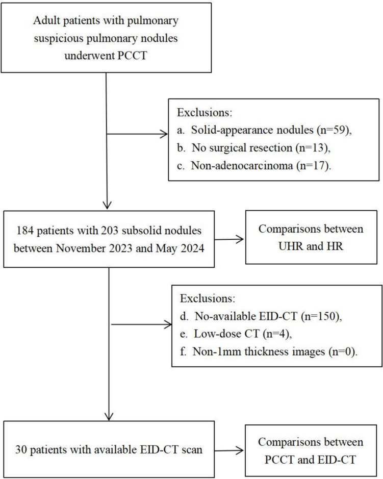
Methods: Patients with subsolid lung adenocarcinoma were recruited for chest scan on PCCT to obtain UHR and standard HR images between November 2023 and May 2024. Nodule characteristics were visually assessed and histogram features were extracted from each nodule. Image quality and radiation dose at previous energy-integrating detector CT (EID-CT) of 30 patients were compared with those of PCCT. Differences between UHR and standard HR, PCCT and EID-CT were compared using paired McNemar-test or paired Wilcox-test.
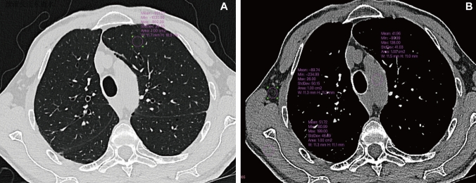
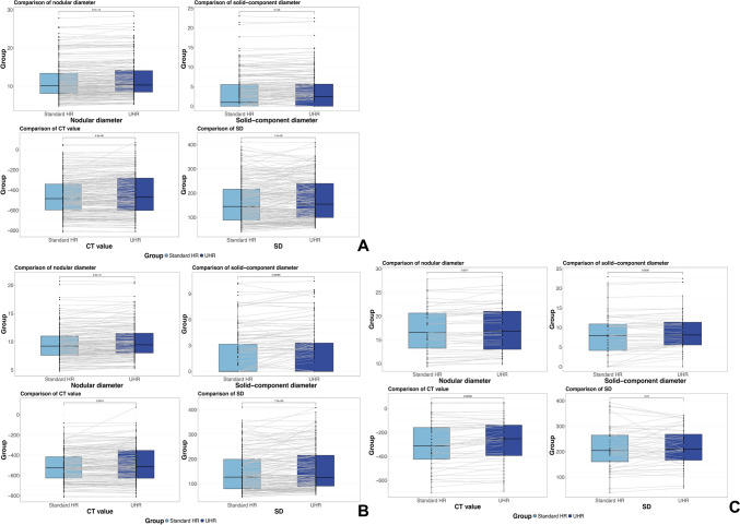
Results: One hundred and eighty-four patients with 203 subsolid nodules were collected including 77 precursors, 77 minimally invasive adenocarcinoma (MIA) and 49 IA. UHR significantly outperformed standard HR in revealing CT findings including larger nodular diameter and solid-component diameter, more frequency of heterogeneous attenuation, lobulation, bubble-like sign, air bronchogram, pleural indentation and vascular sign (all P < 0.05). Additionally, UHR images exhibited significantly greater value in histogram-derived parameters compared to standard HR images (all P < 0.05), except for "Median," "Minimum." Furthermore, the radiation dose in PCCT was half of that in EID-CT (effective dose: 1.32 ± 0.27 vs. 3.85 ± 1.65/mSv, P < 0.001. CDTIvol: 2.97 ± 0.53 vs. 6.90 ± 2.97/mGy, P < 0.001), with image quality significantly better in PCCT.
Conclusion: The UHR protocol on PCCT provides a magnified perspective to reveal CT characteristics of invasive growth in subsolid LUAD, previously undetectable on standard HR images, achieving halved radiation dose and better image quality than EID-CT.
期刊介绍:
Felice Perussia founded La radiologia medica in 1914. It is a peer-reviewed journal and serves as the official journal of the Italian Society of Medical and Interventional Radiology (SIRM). The primary purpose of the journal is to disseminate information related to Radiology, especially advancements in diagnostic imaging and related disciplines. La radiologia medica welcomes original research on both fundamental and clinical aspects of modern radiology, with a particular focus on diagnostic and interventional imaging techniques. It also covers topics such as radiotherapy, nuclear medicine, radiobiology, health physics, and artificial intelligence in the context of clinical implications. The journal includes various types of contributions such as original articles, review articles, editorials, short reports, and letters to the editor. With an esteemed Editorial Board and a selection of insightful reports, the journal is an indispensable resource for radiologists and professionals in related fields. Ultimately, La radiologia medica aims to serve as a platform for international collaboration and knowledge sharing within the radiological community.

 求助内容:
求助内容: 应助结果提醒方式:


