A low-cost, high-throughput pipeline for 3D imaging of embryonic mouse hearts using lightsheet microscopy
IF 2.1
3区 生物学
Q2 DEVELOPMENTAL BIOLOGY
引用次数: 0
Abstract
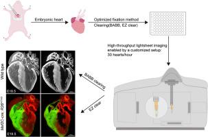
Lightsheet microscopy is a powerful tool for three-dimensional imaging of both live and fixed specimens, spanning small to large scales. However, the time-intensive sample preparation and mounting process often limit its use in high-throughput studies. Given our focus on cardiac development and identifying cardiovascular abnormalities after teratogenic exposure, we have developed an efficient, low-cost, and user-friendly system for specimen fixation, clearing, and mounting. This system enables rapid 3D imaging of a mouse embryonic heart within 1 min using the Zeiss Lightsheet Z.1 microscopy and supports imaging of at least 30 hearts per hour with high resolution. With this system, we obtained high-quality images of embryonic hearts at various stages, visualizing internal structures like the aortic valve and coronary arteries with this system. We further demonstrated its capability for quantitative analysis, including endocardial cushion cell density at E10.5 and volumetric measurements of valve morphology. As an extended application, the system was also applied to postnatal P10 hearts and extra-cardiac organs like kidney and ovary, showing clear structural detail. Additionally, integration of the water-soluble clearing agent, EZ Clear, alongside Cre-loxP-mediated genetic lineage tracing, enabled 3D visualization of cellular contributions to heart development with high resolution. The sample preparation system described here promises broader applications in embryology, anatomy, and pathology research, especially in studies requiring both high throughput and high resolution of 3D imaging.
一种低成本、高通量的胚胎小鼠心脏三维成像通道。
光片显微镜是一个强大的工具,为三维成像的活和固定标本,跨越小到大尺度。然而,耗时的样品制备和安装过程往往限制了其在高通量研究中的使用。鉴于我们专注于心脏发育和识别致畸暴露后的心血管异常,我们开发了一种高效、低成本、用户友好的标本固定、清除和安装系统。该系统可以在一分钟内使用蔡司Lightsheet Z.1显微镜对小鼠胚胎心脏进行快速3D成像,并支持每小时至少30颗心脏的高分辨率成像。通过这个系统,我们获得了胚胎心脏在不同阶段的高质量图像,可视化内部结构,如主动脉瓣和冠状动脉。我们进一步证明了其定量分析的能力,包括E10.5时的心内膜缓冲细胞密度和瓣膜形态的体积测量。作为扩展应用,该系统还应用于出生后P10心脏和肾、卵巢等心外器官,显示出清晰的结构细节。此外,将水溶性清除剂EZ Clear与cre - loxp介导的遗传谱系追踪相结合,能够以高分辨率实现细胞对心脏发育的贡献的3D可视化。本文描述的样品制备系统有望在胚胎学、解剖学和病理学研究中得到更广泛的应用,特别是在需要高通量和高分辨率3D成像的研究中。
本文章由计算机程序翻译,如有差异,请以英文原文为准。
求助全文
约1分钟内获得全文
求助全文
来源期刊
Developmental biology
生物-发育生物学
CiteScore
5.30
自引率
3.70%
发文量
182
审稿时长
1.5 months
期刊介绍:
Developmental Biology (DB) publishes original research on mechanisms of development, differentiation, and growth in animals and plants at the molecular, cellular, genetic and evolutionary levels. Areas of particular emphasis include transcriptional control mechanisms, embryonic patterning, cell-cell interactions, growth factors and signal transduction, and regulatory hierarchies in developing plants and animals.
 求助内容:
求助内容: 应助结果提醒方式:


