Alan Ramalho, Marie-Frédérique Gauthier, Ina Maltais-Payette, Giada Ostinelli, Frédéric Hould, Laurent Biertho, André Tchernof
{"title":"High-throughput measurement of adipocyte size with open-source software using whole-slide adipose tissue images.","authors":"Alan Ramalho, Marie-Frédérique Gauthier, Ina Maltais-Payette, Giada Ostinelli, Frédéric Hould, Laurent Biertho, André Tchernof","doi":"10.1080/21623945.2025.2528437","DOIUrl":null,"url":null,"abstract":"<p><p>The aim of this study was to create and validate a high-throughput method based on open-source software for the measurement of adipocyte diameters in white adipose tissue histological sections. Human omental and subcutaneous adipose tissue samples collected during bariatric surgery were used to prepare haematoxylin and eosin-stained histological slides. Adipocyte diameters were measured both manually and with an automated procedure created using ImageJ. Comparative analysis of our automated method with the manual measurement and associations of the mean adipocyte diameters with cardiometabolic markers were used to validate our method. A total of 377 adipose samples (190 participants) were included in the analysis. Pearson correlation of mean adipocyte diameters showed a strong linear relationship between methods (<i>r</i> = 0.87, <i>p</i> < 0.0001). Omental adipocyte diameters of both methods were significantly associated with the same markers of cardiometabolic risk (fasting concentrations of TG, HDL-Chol, homoeostasis model assessment of insulin resistance, and visceral adiposity index values) with no significant differences between methods. There were also no significant differences between the manual and automated method regarding the correlations between mean subcutaneous adipocyte diameters and anthropometric or metabolic markers. In conclusion, we have created and validated a rapid automated method to measure adipocyte diameters from whole-slide adipose tissue images.</p>","PeriodicalId":7226,"journal":{"name":"Adipocyte","volume":"14 1","pages":"2528437"},"PeriodicalIF":3.1000,"publicationDate":"2025-12-01","publicationTypes":"Journal Article","fieldsOfStudy":null,"isOpenAccess":false,"openAccessPdf":"https://www.ncbi.nlm.nih.gov/pmc/articles/PMC12309532/pdf/","citationCount":"0","resultStr":null,"platform":"Semanticscholar","paperid":null,"PeriodicalName":"Adipocyte","FirstCategoryId":"99","ListUrlMain":"https://doi.org/10.1080/21623945.2025.2528437","RegionNum":4,"RegionCategory":"生物学","ArticlePicture":[],"TitleCN":null,"AbstractTextCN":null,"PMCID":null,"EPubDate":"2025/7/28 0:00:00","PubModel":"Epub","JCR":"Q2","JCRName":"ENDOCRINOLOGY & METABOLISM","Score":null,"Total":0}
引用次数: 0
Abstract
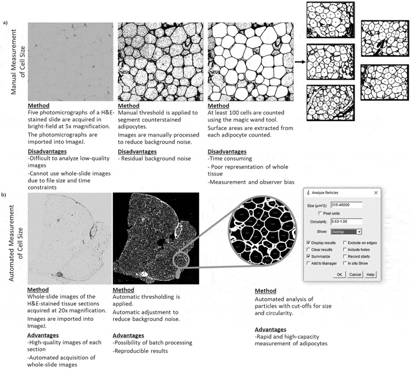
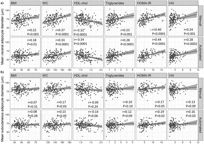
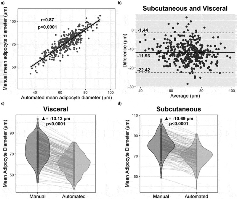
The aim of this study was to create and validate a high-throughput method based on open-source software for the measurement of adipocyte diameters in white adipose tissue histological sections. Human omental and subcutaneous adipose tissue samples collected during bariatric surgery were used to prepare haematoxylin and eosin-stained histological slides. Adipocyte diameters were measured both manually and with an automated procedure created using ImageJ. Comparative analysis of our automated method with the manual measurement and associations of the mean adipocyte diameters with cardiometabolic markers were used to validate our method. A total of 377 adipose samples (190 participants) were included in the analysis. Pearson correlation of mean adipocyte diameters showed a strong linear relationship between methods (r = 0.87, p < 0.0001). Omental adipocyte diameters of both methods were significantly associated with the same markers of cardiometabolic risk (fasting concentrations of TG, HDL-Chol, homoeostasis model assessment of insulin resistance, and visceral adiposity index values) with no significant differences between methods. There were also no significant differences between the manual and automated method regarding the correlations between mean subcutaneous adipocyte diameters and anthropometric or metabolic markers. In conclusion, we have created and validated a rapid automated method to measure adipocyte diameters from whole-slide adipose tissue images.
本研究的目的是创建并验证一种基于开源软件的高通量方法,用于测量白色脂肪组织组织学切片中的脂肪细胞直径。在减肥手术中收集的人网膜和皮下脂肪组织样本用于制备血红素和伊红染色的组织学切片。脂肪细胞直径采用人工和使用ImageJ创建的自动程序测量。将我们的自动化方法与人工测量方法进行比较分析,并将平均脂肪细胞直径与心脏代谢标志物的关联用于验证我们的方法。共有377份脂肪样本(190名参与者)被纳入分析。平均脂肪细胞直径的Pearson相关性显示两种方法之间有很强的线性关系(r = 0.87, p
期刊介绍:
Adipocyte recognizes that the adipose tissue is the largest endocrine organ in the body, and explores the link between dysfunctional adipose tissue and the growing number of chronic diseases including diabetes, hypertension, cardiovascular disease and cancer. Historically, the primary function of the adipose tissue was limited to energy storage and thermoregulation. However, a plethora of research over the past 3 decades has recognized the dynamic role of the adipose tissue and its contribution to a variety of physiological processes including reproduction, angiogenesis, apoptosis, inflammation, blood pressure, coagulation, fibrinolysis, immunity and general metabolic homeostasis. The field of Adipose Tissue research has grown tremendously, and Adipocyte is the first international peer-reviewed journal of its kind providing a multi-disciplinary forum for research focusing exclusively on all aspects of adipose tissue physiology and pathophysiology. Adipocyte accepts high-profile submissions in basic, translational and clinical research.
 求助内容:
求助内容: 应助结果提醒方式:


