{"title":"Fabrication and Characterization of 3D Printed Polycaprolactone/Baghdadite/Zinc Oxide Nanocomposite Scaffolds for Bone Tissue Engineering","authors":"Zahra Safaei, Mojtaba Ansari, Hossein Eslami","doi":"10.1002/bip.70041","DOIUrl":null,"url":null,"abstract":"<div>\n \n <p>Nowadays, the use of 3D printing method in the construction of scaffolds is significantly common for bone tissue engineering applications. Moreover, the addition of nanoparticles and additives can significantly improve the mechanical and biological properties of polymeric scaffolds as polymers alone are not able to show enough performances. In this study, composite scaffolds based on polycaprolactone (PCL) containing different amounts of zinc oxide (ZnO) and Baghdadite (B) nanoparticles were fabricated by 3D printing method as novel combinations for bone tissue engineering. Then, their physical, mechanical, and biological properties were investigated. The scanning electron microscopy (SEM) of the composite showed uniform and porous structures with open porosity. Fourier-transform infrared spectroscopy (FTIR) of the scaffolds confirmed that no reaction occurred between PCL, B, and ZnO nanoparticles during the fabrication of composite scaffolds. The PCL/B/ZnO composite scaffolds showed high compressive strength. They also showed weight loss during 4 weeks, which was related to PCL degradation. The high bioactivity of the composite scaffolds was confirmed by dispersive X-ray analysis (EDS). SEM images showed the formation of calcium phosphate (CaP) layer on scaffolds in simulated body fluid (SBF). Inductively coupled plasma (ICP) analysis confirmed the formation of apatite layer on their surfaces. Based on the results of the (3-(4,5-Dimethylthiazol-2-yl)-2,5-Diphenyltetrazolium Bromide) (MTT) test, cell proliferation on the scaffolds increased after 72 h, which shows that the scaffolds are biocompatible and non-toxic. SEM images showed that the cells on the surface of PCL-based nanocomposite scaffolds prepared had a suitable density. The results of alizarin red staining showed a significant amount of calcium deposition on the scaffolds. It has been shown that PCL-based nanocomposite scaffolds containing B and ZnO nanoparticles are suitable candidates for use in bone tissue engineering applications as they have suitable mechanical, biological, and physical properties.</p>\n </div>","PeriodicalId":8866,"journal":{"name":"Biopolymers","volume":"116 5","pages":""},"PeriodicalIF":3.2000,"publicationDate":"2025-07-30","publicationTypes":"Journal Article","fieldsOfStudy":null,"isOpenAccess":false,"openAccessPdf":"","citationCount":"0","resultStr":null,"platform":"Semanticscholar","paperid":null,"PeriodicalName":"Biopolymers","FirstCategoryId":"99","ListUrlMain":"https://onlinelibrary.wiley.com/doi/10.1002/bip.70041","RegionNum":4,"RegionCategory":"生物学","ArticlePicture":[],"TitleCN":null,"AbstractTextCN":null,"PMCID":null,"EPubDate":"","PubModel":"","JCR":"Q2","JCRName":"BIOCHEMISTRY & MOLECULAR BIOLOGY","Score":null,"Total":0}
引用次数: 0
Abstract
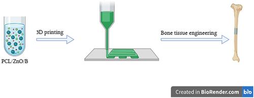
Nowadays, the use of 3D printing method in the construction of scaffolds is significantly common for bone tissue engineering applications. Moreover, the addition of nanoparticles and additives can significantly improve the mechanical and biological properties of polymeric scaffolds as polymers alone are not able to show enough performances. In this study, composite scaffolds based on polycaprolactone (PCL) containing different amounts of zinc oxide (ZnO) and Baghdadite (B) nanoparticles were fabricated by 3D printing method as novel combinations for bone tissue engineering. Then, their physical, mechanical, and biological properties were investigated. The scanning electron microscopy (SEM) of the composite showed uniform and porous structures with open porosity. Fourier-transform infrared spectroscopy (FTIR) of the scaffolds confirmed that no reaction occurred between PCL, B, and ZnO nanoparticles during the fabrication of composite scaffolds. The PCL/B/ZnO composite scaffolds showed high compressive strength. They also showed weight loss during 4 weeks, which was related to PCL degradation. The high bioactivity of the composite scaffolds was confirmed by dispersive X-ray analysis (EDS). SEM images showed the formation of calcium phosphate (CaP) layer on scaffolds in simulated body fluid (SBF). Inductively coupled plasma (ICP) analysis confirmed the formation of apatite layer on their surfaces. Based on the results of the (3-(4,5-Dimethylthiazol-2-yl)-2,5-Diphenyltetrazolium Bromide) (MTT) test, cell proliferation on the scaffolds increased after 72 h, which shows that the scaffolds are biocompatible and non-toxic. SEM images showed that the cells on the surface of PCL-based nanocomposite scaffolds prepared had a suitable density. The results of alizarin red staining showed a significant amount of calcium deposition on the scaffolds. It has been shown that PCL-based nanocomposite scaffolds containing B and ZnO nanoparticles are suitable candidates for use in bone tissue engineering applications as they have suitable mechanical, biological, and physical properties.
期刊介绍:
Founded in 1963, Biopolymers publishes strictly peer-reviewed papers examining naturally occurring and synthetic biological macromolecules. By including experimental and theoretical studies on the fundamental behaviour as well as applications of biopolymers, the journal serves the interdisciplinary biochemical, biophysical, biomaterials and biomedical research communities.
 求助内容:
求助内容: 应助结果提醒方式:


