{"title":"LncRNA LUCAT1 as a prognostic biomarker in cholangiocarcinoma through targeting miR-141-3p: clinical and functional insights.","authors":"Yuxin An, Qing Chen, Shanshan Zhou, Chengcheng Ying, Guanbao Long, Zouxiao Hu, Jiangyang Sun, Niu Zhang","doi":"10.1186/s41065-025-00512-6","DOIUrl":null,"url":null,"abstract":"<p><strong>Background: </strong>Cholangiocarcinoma (CHOL) has a poor prognosis due to its asymptomatic progression, challenges in early detection, and limited treatment options. The lncRNA LUCAT1 is highly expressed in several cancers, including lung, gastric, ovarian, and osteosarcoma tissues.</p><p><strong>Aim: </strong>This study investigates the potential of LUCAT1 as a diagnostic and prognostic biomarker for CHOL.</p><p><strong>Materials and methods: </strong>In this study, we collected tumor tissues and adjacent tumor healthy tissues from 83 CHOL patients. LUCAT1 expression was quantified in CHOL tissues and cell lines via RT-qPCR. Diagnostic and prognostic significance was assessed through ROC curves, Kaplan-Meier survival analysis, and Cox regression models. The biological effects of LUCAT1 on cell proliferation and migration were examined using QBC939 and HuCCT1 cells with transfection assays. The regulatory interaction between LUCAT1 and miR-141-3p was validated using a dual-luciferase reporter assay.</p><p><strong>Results: </strong>Elevated expression of LUCAT1 was observed in CHOL tumor tissues and human cholangiocarcinoma cells, correlating with tumor size, CA-19-9 levels, and TNM stage. The ROC curve, with an AUC of 0.908 (p < 0.001), effectively distinguished CHOL tumor tissues from adjacent non-tumor tissues. And its sensitivity and specificity in distinguishing CHOL tissues from normal tissues were 88.5% and 89.2%, respectively. Survival analyses linked LUCAT1 overexpression to poorer patient outcomes. Silencing LUCAT1 impaired the proliferation and migration of QBC939 and HuCCT1 cells. Dual-luciferase assay confirmed the regulatory relationship between miR-141-3p and LUCAT1. Inhibition of miR-141-3p reversed the effect of LUCAT1 on the proliferation and migration of QBC939 and HuCCT1 cells.</p><p><strong>Conclusion: </strong>LUCAT1 demonstrates significant diagnostic and prognostic potential and could serve as a novel biomarker for CHOL.</p>","PeriodicalId":12862,"journal":{"name":"Hereditas","volume":"162 1","pages":"143"},"PeriodicalIF":2.5000,"publicationDate":"2025-07-26","publicationTypes":"Journal Article","fieldsOfStudy":null,"isOpenAccess":false,"openAccessPdf":"https://www.ncbi.nlm.nih.gov/pmc/articles/PMC12297753/pdf/","citationCount":"0","resultStr":null,"platform":"Semanticscholar","paperid":null,"PeriodicalName":"Hereditas","FirstCategoryId":"99","ListUrlMain":"https://doi.org/10.1186/s41065-025-00512-6","RegionNum":3,"RegionCategory":"生物学","ArticlePicture":[],"TitleCN":null,"AbstractTextCN":null,"PMCID":null,"EPubDate":"","PubModel":"","JCR":"","JCRName":"","Score":null,"Total":0}
引用次数: 0
Abstract
Background: Cholangiocarcinoma (CHOL) has a poor prognosis due to its asymptomatic progression, challenges in early detection, and limited treatment options. The lncRNA LUCAT1 is highly expressed in several cancers, including lung, gastric, ovarian, and osteosarcoma tissues.
Aim: This study investigates the potential of LUCAT1 as a diagnostic and prognostic biomarker for CHOL.
Materials and methods: In this study, we collected tumor tissues and adjacent tumor healthy tissues from 83 CHOL patients. LUCAT1 expression was quantified in CHOL tissues and cell lines via RT-qPCR. Diagnostic and prognostic significance was assessed through ROC curves, Kaplan-Meier survival analysis, and Cox regression models. The biological effects of LUCAT1 on cell proliferation and migration were examined using QBC939 and HuCCT1 cells with transfection assays. The regulatory interaction between LUCAT1 and miR-141-3p was validated using a dual-luciferase reporter assay.
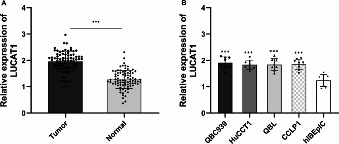
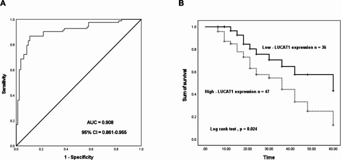
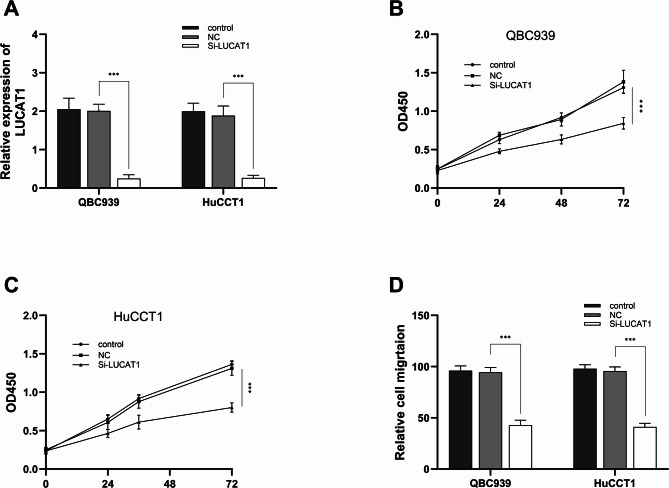
Results: Elevated expression of LUCAT1 was observed in CHOL tumor tissues and human cholangiocarcinoma cells, correlating with tumor size, CA-19-9 levels, and TNM stage. The ROC curve, with an AUC of 0.908 (p < 0.001), effectively distinguished CHOL tumor tissues from adjacent non-tumor tissues. And its sensitivity and specificity in distinguishing CHOL tissues from normal tissues were 88.5% and 89.2%, respectively. Survival analyses linked LUCAT1 overexpression to poorer patient outcomes. Silencing LUCAT1 impaired the proliferation and migration of QBC939 and HuCCT1 cells. Dual-luciferase assay confirmed the regulatory relationship between miR-141-3p and LUCAT1. Inhibition of miR-141-3p reversed the effect of LUCAT1 on the proliferation and migration of QBC939 and HuCCT1 cells.
Conclusion: LUCAT1 demonstrates significant diagnostic and prognostic potential and could serve as a novel biomarker for CHOL.
HereditasBiochemistry, Genetics and Molecular Biology-Genetics
CiteScore
3.80
自引率
3.70%
发文量
0
期刊介绍:
For almost a century, Hereditas has published original cutting-edge research and reviews. As the Official journal of the Mendelian Society of Lund, the journal welcomes research from across all areas of genetics and genomics. Topics of interest include human and medical genetics, animal and plant genetics, microbial genetics, agriculture and bioinformatics.

 求助内容:
求助内容: 应助结果提醒方式:


