Ran Meng, Bin Huang, Fan Yang, Nannan Zhang, Bin Feng, Dalong Zhu
{"title":"PU.1 Facilitates Endothelial-to-Mesenchymal Transition in Cardiac Endothelial Cells","authors":"Ran Meng, Bin Huang, Fan Yang, Nannan Zhang, Bin Feng, Dalong Zhu","doi":"10.1111/boc.70029","DOIUrl":null,"url":null,"abstract":"<div>\n \n <p><b>Background</b>: The endothelial-to-mesenchymal transition (EndMT) plays a critical role in cardiac fibrosis pathogenesis. However, the molecular mechanisms driving EndMT remain poorly understood. This study investigates the regulatory function of the transcription factor PU.1 in EndMT using primary cardiac endothelial cells.</p>\n <p><b>Methods</b>: Immunofluorescence was performed to assess characteristic protein markers in cultured cells. PU.1 knockdown was achieved through siRNA transfection. Key gene expression was quantified at mRNA and protein levels. EndMT progression was evaluated via migration and tube formation assays. Additionally, immunoprecipitation was utilized to examine PU.1 interaction with phosphorylated Smad3 (p-Smad3).</p>\n <p><b>Results</b>: TGF-β1-induced EndMT is coupled with a significant upregulation of PU.1 expression. PU.1 silencing attenuated EndMT, evidenced by elevated CD31/VE-cadherin and reduced α-SMA/N-cadherin/FSP-1 levels under TGF-β1 stimulation. PU.1 knockdown functionally impaired cell migration while promoting vascular lumenogenesis. Conversely, forced PU.1 expression was sufficient to drive EndMT in cardiac endothelial cells. Mechanistically, our data suggest that PU.1 enhances Smad3 phosphorylation, potentially through direct binding to and stabilization of the p-Smad3 protein.</p>\n <p><b>Conclusion</b>: PU.1 drives EndMT in cardiac endothelial cells by enhancing Smad3 phosphorylation and stability. These results elucidate novel molecular pathways in EndMT and identify PU.1 as a potential therapeutic target for attenuating cardiac fibrosis.</p>\n </div>","PeriodicalId":8859,"journal":{"name":"Biology of the Cell","volume":"117 7","pages":""},"PeriodicalIF":2.4000,"publicationDate":"2025-07-28","publicationTypes":"Journal Article","fieldsOfStudy":null,"isOpenAccess":false,"openAccessPdf":"","citationCount":"0","resultStr":null,"platform":"Semanticscholar","paperid":null,"PeriodicalName":"Biology of the Cell","FirstCategoryId":"99","ListUrlMain":"https://onlinelibrary.wiley.com/doi/10.1111/boc.70029","RegionNum":4,"RegionCategory":"生物学","ArticlePicture":[],"TitleCN":null,"AbstractTextCN":null,"PMCID":null,"EPubDate":"","PubModel":"","JCR":"Q4","JCRName":"CELL BIOLOGY","Score":null,"Total":0}
引用次数: 0
Abstract
Background: The endothelial-to-mesenchymal transition (EndMT) plays a critical role in cardiac fibrosis pathogenesis. However, the molecular mechanisms driving EndMT remain poorly understood. This study investigates the regulatory function of the transcription factor PU.1 in EndMT using primary cardiac endothelial cells.
Methods: Immunofluorescence was performed to assess characteristic protein markers in cultured cells. PU.1 knockdown was achieved through siRNA transfection. Key gene expression was quantified at mRNA and protein levels. EndMT progression was evaluated via migration and tube formation assays. Additionally, immunoprecipitation was utilized to examine PU.1 interaction with phosphorylated Smad3 (p-Smad3).
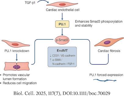
Results: TGF-β1-induced EndMT is coupled with a significant upregulation of PU.1 expression. PU.1 silencing attenuated EndMT, evidenced by elevated CD31/VE-cadherin and reduced α-SMA/N-cadherin/FSP-1 levels under TGF-β1 stimulation. PU.1 knockdown functionally impaired cell migration while promoting vascular lumenogenesis. Conversely, forced PU.1 expression was sufficient to drive EndMT in cardiac endothelial cells. Mechanistically, our data suggest that PU.1 enhances Smad3 phosphorylation, potentially through direct binding to and stabilization of the p-Smad3 protein.
Conclusion: PU.1 drives EndMT in cardiac endothelial cells by enhancing Smad3 phosphorylation and stability. These results elucidate novel molecular pathways in EndMT and identify PU.1 as a potential therapeutic target for attenuating cardiac fibrosis.
期刊介绍:
The journal publishes original research articles and reviews on all aspects of cellular, molecular and structural biology, developmental biology, cell physiology and evolution. It will publish articles or reviews contributing to the understanding of the elementary biochemical and biophysical principles of live matter organization from the molecular, cellular and tissues scales and organisms.
This includes contributions directed towards understanding biochemical and biophysical mechanisms, structure-function relationships with respect to basic cell and tissue functions, development, development/evolution relationship, morphogenesis, stem cell biology, cell biology of disease, plant cell biology, as well as contributions directed toward understanding integrated processes at the organelles, cell and tissue levels. Contributions using approaches such as high resolution imaging, live imaging, quantitative cell biology and integrated biology; as well as those using innovative genetic and epigenetic technologies, ex-vivo tissue engineering, cellular, tissue and integrated functional analysis, and quantitative biology and modeling to demonstrate original biological principles are encouraged.
 求助内容:
求助内容: 应助结果提醒方式:


