Salla Autti, Pauliina Hirvi, Mariia Keitaanniemi, Hanna Mustaniemi, Kalle Kotilahti, Hanna Renvall, Ilkka Nissilä
{"title":"Simultaneously Acquired Magnetoencephalography and Diffuse Optical Tomography Data Reveals Correlated Somatosensory Activity","authors":"Salla Autti, Pauliina Hirvi, Mariia Keitaanniemi, Hanna Mustaniemi, Kalle Kotilahti, Hanna Renvall, Ilkka Nissilä","doi":"10.1002/hbm.70293","DOIUrl":null,"url":null,"abstract":"<p>Simultaneous measurement of electrophysiological and hemodynamic brain signals imposes special requirements on the instrumentation. Here, we developed a high-density fiberoptic probe for concurrent diffuse optical tomography (DOT) and magnetoencephalography (MEG) recordings. Transparent two-component silicone was mixed with carbon black dye to achieve a black, flexible, non-magnetic support for the dense optode arrangement and low (5 mm) probe thickness. The probe was used to record somatosensory responses to electrical right median nerve stimulation at 0.5, 1, 2, and 4 Hz in 18 adult human subjects. Brain activity was simultaneously measured with a commercial whole-head MEG system and with the DOT optode arrangement covering approximately 40 cm<sup>2</sup> over the parietal region in the contralateral left hemisphere. Two correlation-based clustering methods were developed to find regions where the reconstructed time course of total hemoglobin concentration (HbT) changes correlated with the predicted hemodynamic activity based on time-course characteristics of the MEG sources and the canonical hemodynamic response model. Two statistically significant clusters were found based on the correlation between HbT around the postcentral gyrus and MEG primary somatosensory cortical activity at ~35 ms (P35m response). In addition, correlation between HbT and secondary somatosensory cortical activity suggested a statistically significant cluster in the postcentral gyrus and parietal operculum. These results illustrate an improvement in localization over previous DOT studies using sparse optode arrangements, and demonstrate the feasibility of the system for simultaneous HD-DOT-MEG experiments. Furthermore, the techniques described here pave the way for understanding the coupling between hemodynamic and electrophysiological responses. Further research is needed to reveal the neuronal circuits giving rise to the correlating MEG and DOT response features. Significant improvements in the technology are still expected via optimization of the detected light power in the instrumentation.</p>","PeriodicalId":13019,"journal":{"name":"Human Brain Mapping","volume":"46 11","pages":""},"PeriodicalIF":3.3000,"publicationDate":"2025-07-27","publicationTypes":"Journal Article","fieldsOfStudy":null,"isOpenAccess":false,"openAccessPdf":"https://onlinelibrary.wiley.com/doi/epdf/10.1002/hbm.70293","citationCount":"0","resultStr":null,"platform":"Semanticscholar","paperid":null,"PeriodicalName":"Human Brain Mapping","FirstCategoryId":"3","ListUrlMain":"https://onlinelibrary.wiley.com/doi/10.1002/hbm.70293","RegionNum":2,"RegionCategory":"医学","ArticlePicture":[],"TitleCN":null,"AbstractTextCN":null,"PMCID":null,"EPubDate":"","PubModel":"","JCR":"Q1","JCRName":"NEUROIMAGING","Score":null,"Total":0}
引用次数: 0
Abstract
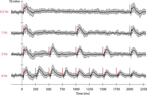
Simultaneous measurement of electrophysiological and hemodynamic brain signals imposes special requirements on the instrumentation. Here, we developed a high-density fiberoptic probe for concurrent diffuse optical tomography (DOT) and magnetoencephalography (MEG) recordings. Transparent two-component silicone was mixed with carbon black dye to achieve a black, flexible, non-magnetic support for the dense optode arrangement and low (5 mm) probe thickness. The probe was used to record somatosensory responses to electrical right median nerve stimulation at 0.5, 1, 2, and 4 Hz in 18 adult human subjects. Brain activity was simultaneously measured with a commercial whole-head MEG system and with the DOT optode arrangement covering approximately 40 cm2 over the parietal region in the contralateral left hemisphere. Two correlation-based clustering methods were developed to find regions where the reconstructed time course of total hemoglobin concentration (HbT) changes correlated with the predicted hemodynamic activity based on time-course characteristics of the MEG sources and the canonical hemodynamic response model. Two statistically significant clusters were found based on the correlation between HbT around the postcentral gyrus and MEG primary somatosensory cortical activity at ~35 ms (P35m response). In addition, correlation between HbT and secondary somatosensory cortical activity suggested a statistically significant cluster in the postcentral gyrus and parietal operculum. These results illustrate an improvement in localization over previous DOT studies using sparse optode arrangements, and demonstrate the feasibility of the system for simultaneous HD-DOT-MEG experiments. Furthermore, the techniques described here pave the way for understanding the coupling between hemodynamic and electrophysiological responses. Further research is needed to reveal the neuronal circuits giving rise to the correlating MEG and DOT response features. Significant improvements in the technology are still expected via optimization of the detected light power in the instrumentation.
同时测量脑电生理和血流动力学信号对仪器有特殊要求。在这里,我们开发了一种高密度光纤探头,用于同时进行漫射光学断层扫描(DOT)和脑磁图(MEG)记录。透明的双组分有机硅与炭黑染料混合,为密集的光电器件排列和低(5毫米)探头厚度提供了黑色、柔性、非磁性的支撑。该探针用于记录18名成人右正中神经0.5、1、2和4 Hz电刺激的体感觉反应。脑活动测量同时使用商用全头部脑磁图系统和DOT光电装置覆盖约40平方厘米的顶叶区域在对侧左半球。基于MEG源的时间过程特征和典型血流动力学反应模型,建立了两种基于相关聚类的方法,寻找重构总血红蛋白浓度(HbT)变化时间过程与预测血流动力学活性相关的区域。基于中枢后回周围HbT与MEG初级体感觉皮层活动在~35 ms (P35m反应)之间的相关性,发现了两个具有统计学意义的簇。此外,HbT与次级体感觉皮层活动之间的相关性表明,在中央后回和顶盖中有统计学意义的集群。这些结果表明,与以往使用稀疏光电器件布置的DOT研究相比,该系统在定位方面有了改进,并证明了该系统用于同时进行HD-DOT-MEG实验的可行性。此外,这里描述的技术为理解血流动力学和电生理反应之间的耦合铺平了道路。进一步的研究需要揭示产生相关脑磁图和DOT响应特征的神经元回路。通过优化仪器中检测到的光功率,该技术仍有望取得重大进展。
期刊介绍:
Human Brain Mapping publishes peer-reviewed basic, clinical, technical, and theoretical research in the interdisciplinary and rapidly expanding field of human brain mapping. The journal features research derived from non-invasive brain imaging modalities used to explore the spatial and temporal organization of the neural systems supporting human behavior. Imaging modalities of interest include positron emission tomography, event-related potentials, electro-and magnetoencephalography, magnetic resonance imaging, and single-photon emission tomography. Brain mapping research in both normal and clinical populations is encouraged.
Article formats include Research Articles, Review Articles, Clinical Case Studies, and Technique, as well as Technological Developments, Theoretical Articles, and Synthetic Reviews. Technical advances, such as novel brain imaging methods, analyses for detecting or localizing neural activity, synergistic uses of multiple imaging modalities, and strategies for the design of behavioral paradigms and neural-systems modeling are of particular interest. The journal endorses the propagation of methodological standards and encourages database development in the field of human brain mapping.

 求助内容:
求助内容: 应助结果提醒方式:


