{"title":"Fractal analysis of mandibular bone structure in individuals with unilateral crossbite.","authors":"Serdar Cik, Gozde Acıkgoz, Ali Kiki","doi":"10.4041/kjod24.296","DOIUrl":null,"url":null,"abstract":"<p><strong>Objective: </strong>This study aimed to examine the effects of unilateral crossbite on the structure of the mandibular bone by using fractal analysis.</p><p><strong>Methods: </strong>This study was conducted using panoramic films obtained retrospectively from 45 individuals with crossbite and 45 individuals with normal occlusion (NO). Fractal measurements were performed on the condyle, corpus, and angulus on both left and right sides of the panoramic films. The films were divided into three groups: cross-side (Cs), normal-side (Ns), and NO. The Cs group was further analyzed on the basis of the number of teeth in the crossbite. Data were analyzed using the non-parametric Kruskal-Wallis test and post-hoc Dunnett's T3 multiple-comparison test. Statistical significance was set at <i>P</i> < 0.05.</p><p><strong>Results: </strong>The mean condylar fractal dimension (FD) showed no significant difference between the groups (<i>P</i> > 0.05). The mean angulus FD in the Cs group was significantly higher than that in the NO group (<i>P</i> = 0.006). Similarly, the mean corpus FD in the NO group was significantly lower than those in the Cs and Ns groups (<i>P</i> = 0.003). In the Cs group, comparisons based on the number of teeth in the crossbite showed no significant differences among the condyle, angulus, or corpus regions.</p><p><strong>Conclusions: </strong>Fractal analysis may be an effective approach for detecting variations in mandibular trabecular patterns associated with unilateral crossbites. In cases of unilateral crossbite, the trabecular structure was affected in the angulus and corpus regions.</p>","PeriodicalId":51260,"journal":{"name":"Korean Journal of Orthodontics","volume":"55 4","pages":"306-313"},"PeriodicalIF":2.3000,"publicationDate":"2025-07-25","publicationTypes":"Journal Article","fieldsOfStudy":null,"isOpenAccess":false,"openAccessPdf":"https://www.ncbi.nlm.nih.gov/pmc/articles/PMC12301423/pdf/","citationCount":"0","resultStr":null,"platform":"Semanticscholar","paperid":null,"PeriodicalName":"Korean Journal of Orthodontics","FirstCategoryId":"3","ListUrlMain":"https://doi.org/10.4041/kjod24.296","RegionNum":3,"RegionCategory":"医学","ArticlePicture":[],"TitleCN":null,"AbstractTextCN":null,"PMCID":null,"EPubDate":"2025/4/30 0:00:00","PubModel":"Epub","JCR":"Q1","JCRName":"DENTISTRY, ORAL SURGERY & MEDICINE","Score":null,"Total":0}
引用次数: 0
Abstract
Objective: This study aimed to examine the effects of unilateral crossbite on the structure of the mandibular bone by using fractal analysis.
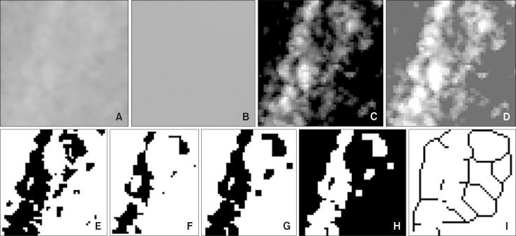
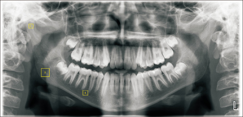
Methods: This study was conducted using panoramic films obtained retrospectively from 45 individuals with crossbite and 45 individuals with normal occlusion (NO). Fractal measurements were performed on the condyle, corpus, and angulus on both left and right sides of the panoramic films. The films were divided into three groups: cross-side (Cs), normal-side (Ns), and NO. The Cs group was further analyzed on the basis of the number of teeth in the crossbite. Data were analyzed using the non-parametric Kruskal-Wallis test and post-hoc Dunnett's T3 multiple-comparison test. Statistical significance was set at P < 0.05.
Results: The mean condylar fractal dimension (FD) showed no significant difference between the groups (P > 0.05). The mean angulus FD in the Cs group was significantly higher than that in the NO group (P = 0.006). Similarly, the mean corpus FD in the NO group was significantly lower than those in the Cs and Ns groups (P = 0.003). In the Cs group, comparisons based on the number of teeth in the crossbite showed no significant differences among the condyle, angulus, or corpus regions.
Conclusions: Fractal analysis may be an effective approach for detecting variations in mandibular trabecular patterns associated with unilateral crossbites. In cases of unilateral crossbite, the trabecular structure was affected in the angulus and corpus regions.
期刊介绍:
The Korean Journal of Orthodontics (KJO) is an international, open access, peer reviewed journal published in January, March, May, July, September, and November each year. It was first launched in 1970 and, as the official scientific publication of Korean Association of Orthodontists, KJO aims to publish high quality clinical and scientific original research papers in all areas related to orthodontics and dentofacial orthopedics. Specifically, its interest focuses on evidence-based investigations of contemporary diagnostic procedures and treatment techniques, expanding to significant clinical reports of diverse treatment approaches.
The scope of KJO covers all areas of orthodontics and dentofacial orthopedics including successful diagnostic procedures and treatment planning, growth and development of the face and its clinical implications, appliance designs, biomechanics, TMJ disorders and adult treatment. Specifically, its latest interest focuses on skeletal anchorage devices, orthodontic appliance and biomaterials, 3 dimensional imaging techniques utilized for dentofacial diagnosis and treatment planning, and orthognathic surgery to correct skeletal disharmony in association of orthodontic treatment.
 求助内容:
求助内容: 应助结果提醒方式:


