Mechanism of 15-hydroxyprostaglandin dehydrogenase protein inhibiting cervical cancer cell proliferation through downregulation of the notch1 signaling pathway.
{"title":"Mechanism of 15-hydroxyprostaglandin dehydrogenase protein inhibiting cervical cancer cell proliferation through downregulation of the notch1 signaling pathway.","authors":"Suwen Chang","doi":"10.25259/Cytojournal_57_2025","DOIUrl":null,"url":null,"abstract":"<p><strong>Objective: </strong>This study aims to explore the modulatory effect of 15-hydroxyprostaglandin dehydrogenase (15-PGDH) protein in the Notch1 signaling pathway in cervical cancer (CC) cells and assess how this modulation affects the proliferation and migration of CC cells. Moreover, this study offers fresh perspectives on the molecular mechanisms underlying CC by thoroughly analyzing the relationship between 15-PGDH and the Notch1 signaling pathway, and investigates the therapeutic potential of 15-PGDH.</p><p><strong>Material and methods: </strong>Human normal cervical epithelial cells and CC cell lines (human CC cell line [HeLa], human cervical squamous carcinoma cell line [Caski], and human cervical epidermoid carcinoma cells [ME180]) were selected as experimental models. Western blotting (WB) and quantitative reverse transcription polymerase chain reaction were performed to evaluate the protein and messenger RNA levels of 15-PGDH and Notch receptor 1 (Notch1) signaling pathway-related proteins (Jagged canonical Notch ligand 1 [Jagged1] and Hes family bHLH transcription factor 1 [Hes1]). Results suggested that the HeLa and Caski cells exhibited significant expression of 15-PGDH and Notch1 signaling-related proteins. A series of experiments, including WB, cell counting kit-8 assay, Transwell migration assay, and 5-ethynyl-2'-deoxyuridine assay, was conducted in the HeLa and Caski cells to obtain an extensive understanding of how 15-PGDH influences Notch1 signaling regulation. This study also utilized the 15-PGDH inhibitor SW033291 and a Notch1 overexpression vector to evaluate the effect of 15-PGDH on CC cell growth, motility, and Notch1 signaling pathway modulation.</p><p><strong>Results: </strong>Results demonstrated that in the normal human cervical epithelial cells, 15-PGDH was highly expressed, while the Notch1 signaling pathway-related proteins exhibited low expression quantities. However, in HeLa and Caski CC cells, 15-PGDH expression was significantly downregulated (<i>P</i> < 0.001 or <i>P</i> < 0.01), whereas the Notch1 signaling pathway was activated. Further studies revealed that 15-PGDH or its inhibitor influenced the stimulation of the Notch1 signaling pathway in the HeLa and Caski cells. Specifically, the 15-PGDH inhibitor SW033291 reduced 15-PGDH expression (<i>P</i> < 0.001 or <i>P</i> < 0.01) and promoted Notch signaling activation. Meanwhile, 15-PGDH upregulation suppressed Notch signaling activation. Furthermore, 15-PGDH successfully prevented the proliferation and migration of CC cells induced by Notch1 overexpression and reduced the activation of the Notch signaling pathway, as shown by the downregulation of Notch1 and its downstream effectors, Jagged1 and Hes1.</p><p><strong>Conclusion: </strong>This study highlights the role of 15-PGDH in regulating the Notch1 signaling pathway in CC cells, focusing on its effect on cell proliferation and migration. The results demonstrate that 15-PGDH suppresses CC cell proliferation and migration by downregulating the Notch1 signaling pathway. These findings provide new insights into the molecular mechanisms underlying CC and suggest 15-PGDH as a promising therapeutic target.</p>","PeriodicalId":49082,"journal":{"name":"Cytojournal","volume":"22 ","pages":"59"},"PeriodicalIF":3.1000,"publicationDate":"2025-06-13","publicationTypes":"Journal Article","fieldsOfStudy":null,"isOpenAccess":false,"openAccessPdf":"https://www.ncbi.nlm.nih.gov/pmc/articles/PMC12289113/pdf/","citationCount":"0","resultStr":null,"platform":"Semanticscholar","paperid":null,"PeriodicalName":"Cytojournal","FirstCategoryId":"3","ListUrlMain":"https://doi.org/10.25259/Cytojournal_57_2025","RegionNum":4,"RegionCategory":"医学","ArticlePicture":[],"TitleCN":null,"AbstractTextCN":null,"PMCID":null,"EPubDate":"2025/1/1 0:00:00","PubModel":"eCollection","JCR":"Q2","JCRName":"PATHOLOGY","Score":null,"Total":0}
引用次数: 0
Abstract
Objective: This study aims to explore the modulatory effect of 15-hydroxyprostaglandin dehydrogenase (15-PGDH) protein in the Notch1 signaling pathway in cervical cancer (CC) cells and assess how this modulation affects the proliferation and migration of CC cells. Moreover, this study offers fresh perspectives on the molecular mechanisms underlying CC by thoroughly analyzing the relationship between 15-PGDH and the Notch1 signaling pathway, and investigates the therapeutic potential of 15-PGDH.
Material and methods: Human normal cervical epithelial cells and CC cell lines (human CC cell line [HeLa], human cervical squamous carcinoma cell line [Caski], and human cervical epidermoid carcinoma cells [ME180]) were selected as experimental models. Western blotting (WB) and quantitative reverse transcription polymerase chain reaction were performed to evaluate the protein and messenger RNA levels of 15-PGDH and Notch receptor 1 (Notch1) signaling pathway-related proteins (Jagged canonical Notch ligand 1 [Jagged1] and Hes family bHLH transcription factor 1 [Hes1]). Results suggested that the HeLa and Caski cells exhibited significant expression of 15-PGDH and Notch1 signaling-related proteins. A series of experiments, including WB, cell counting kit-8 assay, Transwell migration assay, and 5-ethynyl-2'-deoxyuridine assay, was conducted in the HeLa and Caski cells to obtain an extensive understanding of how 15-PGDH influences Notch1 signaling regulation. This study also utilized the 15-PGDH inhibitor SW033291 and a Notch1 overexpression vector to evaluate the effect of 15-PGDH on CC cell growth, motility, and Notch1 signaling pathway modulation.
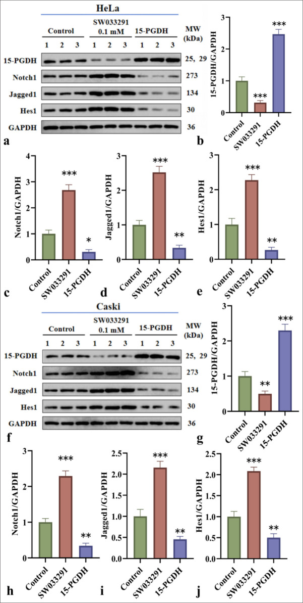
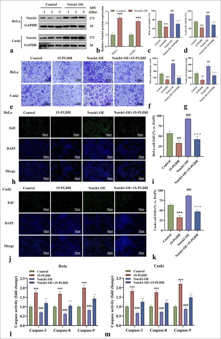
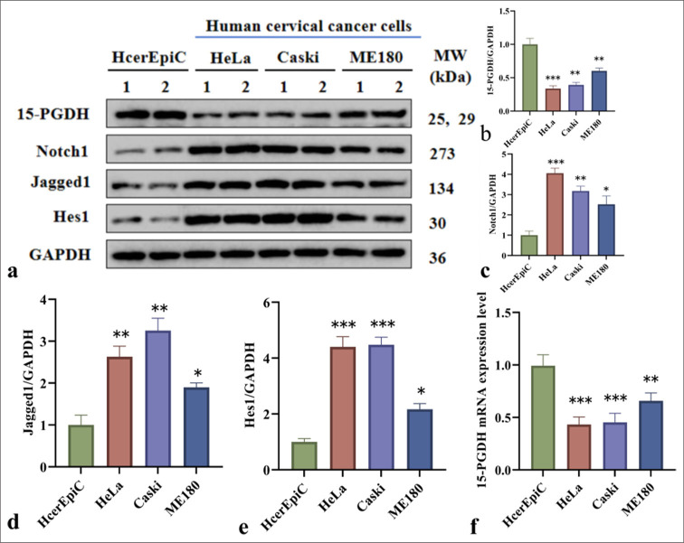
Results: Results demonstrated that in the normal human cervical epithelial cells, 15-PGDH was highly expressed, while the Notch1 signaling pathway-related proteins exhibited low expression quantities. However, in HeLa and Caski CC cells, 15-PGDH expression was significantly downregulated (P < 0.001 or P < 0.01), whereas the Notch1 signaling pathway was activated. Further studies revealed that 15-PGDH or its inhibitor influenced the stimulation of the Notch1 signaling pathway in the HeLa and Caski cells. Specifically, the 15-PGDH inhibitor SW033291 reduced 15-PGDH expression (P < 0.001 or P < 0.01) and promoted Notch signaling activation. Meanwhile, 15-PGDH upregulation suppressed Notch signaling activation. Furthermore, 15-PGDH successfully prevented the proliferation and migration of CC cells induced by Notch1 overexpression and reduced the activation of the Notch signaling pathway, as shown by the downregulation of Notch1 and its downstream effectors, Jagged1 and Hes1.
Conclusion: This study highlights the role of 15-PGDH in regulating the Notch1 signaling pathway in CC cells, focusing on its effect on cell proliferation and migration. The results demonstrate that 15-PGDH suppresses CC cell proliferation and migration by downregulating the Notch1 signaling pathway. These findings provide new insights into the molecular mechanisms underlying CC and suggest 15-PGDH as a promising therapeutic target.
期刊介绍:
The CytoJournal is an open-access peer-reviewed journal committed to publishing high-quality articles in the field of Diagnostic Cytopathology including Molecular aspects. The journal is owned by the Cytopathology Foundation and published by the Scientific Scholar.
 求助内容:
求助内容: 应助结果提醒方式:


