{"title":"Endoscopic ultrasound-guided fine-needle aspiration value in suspected autoimmune pancreatitis malignancy diagnosis.","authors":"Yue Liu, Dongling Wan, Chang Wu, Deyu Zhang, Jiaheng Xu, Wanshun Li, Zhenghui Yang, Jiayu Li, Ying Chen, Zhendong Jin, Haojie Huang","doi":"10.25259/Cytojournal_214_2024","DOIUrl":null,"url":null,"abstract":"<p><strong>Objective: </strong>Histopathology examination is important for diagnosing autoimmune pancreatitis (AIP), which is suspected to be pancreatic cancer based on imaging findings. Although the validity of endoscopic ultrasound-guided fine-needle aspiration (EUS-FNA) in the diagnosis of AIP is still debated globally, this study aimed to evaluate the efficacy of EUS-FNA in the diagnosis of AIP with suspected pancreatic cancer.</p><p><strong>Material and methods: </strong>From January 2021 to June 2024, 30 AIP patients with radiographically diagnosed pancreatic cancer were enrolled and underwent EUS-FNA. Sex, age, symptoms, CA199, serum immunoglobulin G4 (IgG4), and treatment outcome were included. Tissue sampling conditions, puncture sites, storiform fibrosis, CD38- and IgG4-positive plasma cell counts, and obliterans phlebitis were evaluated.</p><p><strong>Results: </strong>Thirty patients, 24 males and six females, with an average age of 60.53 ± 11.72 years (32-79 years), were included in the study. Thirty patients had their serum IgG4 and CA199 levels tested. Tissue samples containing ≥10 were obtained from 19 (63.33%) patients. CD38+ plasma cell infiltration and laminar fibrosis were detected in 22 (73.33%) and 10 (33.33%) patients. According to the International Consensus Diagnostic Criteria ( ICDC), 12 patients had histopathological levels of Grade 1, 15 of Grade 2, and three patients could not be classified. The accuracy, sensitivity, and specificity of EUS-FNA in diagnosing AIP with suspected pancreatic cancer on imaging were 96.66% (29/30), 96.42% (27/28), and 100% (2/2), respectively. The area under the curve value of EUS-FNA for patients with AIP who were radiologically suspected of having pancreatic cancer was 0.957.</p><p><strong>Conclusion: </strong>Approximately 90% of patients with EUS-FNA results are diagnosed with an ICDC level of 2 or higher. Our results suggest that for cases where malignant tumors are suspected after imaging or cannot be ruled out, obtaining pancreatic tissue through EUS-FNA puncture for pathological diagnosis is recommended.</p>","PeriodicalId":49082,"journal":{"name":"Cytojournal","volume":"22 ","pages":"58"},"PeriodicalIF":3.1000,"publicationDate":"2025-06-02","publicationTypes":"Journal Article","fieldsOfStudy":null,"isOpenAccess":false,"openAccessPdf":"https://www.ncbi.nlm.nih.gov/pmc/articles/PMC12289111/pdf/","citationCount":"0","resultStr":null,"platform":"Semanticscholar","paperid":null,"PeriodicalName":"Cytojournal","FirstCategoryId":"3","ListUrlMain":"https://doi.org/10.25259/Cytojournal_214_2024","RegionNum":4,"RegionCategory":"医学","ArticlePicture":[],"TitleCN":null,"AbstractTextCN":null,"PMCID":null,"EPubDate":"2025/1/1 0:00:00","PubModel":"eCollection","JCR":"Q2","JCRName":"PATHOLOGY","Score":null,"Total":0}
引用次数: 0
Abstract
Objective: Histopathology examination is important for diagnosing autoimmune pancreatitis (AIP), which is suspected to be pancreatic cancer based on imaging findings. Although the validity of endoscopic ultrasound-guided fine-needle aspiration (EUS-FNA) in the diagnosis of AIP is still debated globally, this study aimed to evaluate the efficacy of EUS-FNA in the diagnosis of AIP with suspected pancreatic cancer.
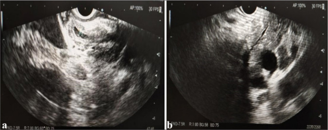
Material and methods: From January 2021 to June 2024, 30 AIP patients with radiographically diagnosed pancreatic cancer were enrolled and underwent EUS-FNA. Sex, age, symptoms, CA199, serum immunoglobulin G4 (IgG4), and treatment outcome were included. Tissue sampling conditions, puncture sites, storiform fibrosis, CD38- and IgG4-positive plasma cell counts, and obliterans phlebitis were evaluated.
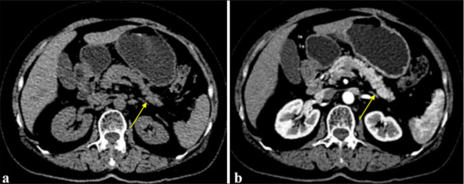
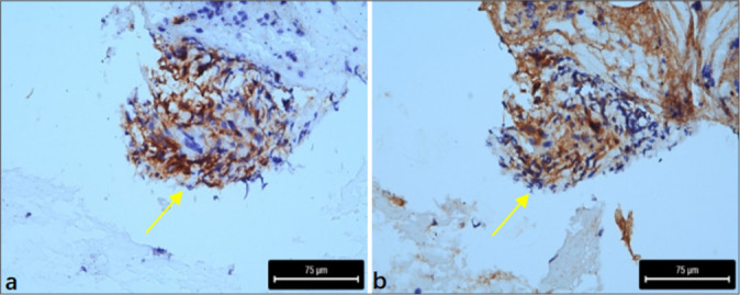
Results: Thirty patients, 24 males and six females, with an average age of 60.53 ± 11.72 years (32-79 years), were included in the study. Thirty patients had their serum IgG4 and CA199 levels tested. Tissue samples containing ≥10 were obtained from 19 (63.33%) patients. CD38+ plasma cell infiltration and laminar fibrosis were detected in 22 (73.33%) and 10 (33.33%) patients. According to the International Consensus Diagnostic Criteria ( ICDC), 12 patients had histopathological levels of Grade 1, 15 of Grade 2, and three patients could not be classified. The accuracy, sensitivity, and specificity of EUS-FNA in diagnosing AIP with suspected pancreatic cancer on imaging were 96.66% (29/30), 96.42% (27/28), and 100% (2/2), respectively. The area under the curve value of EUS-FNA for patients with AIP who were radiologically suspected of having pancreatic cancer was 0.957.
Conclusion: Approximately 90% of patients with EUS-FNA results are diagnosed with an ICDC level of 2 or higher. Our results suggest that for cases where malignant tumors are suspected after imaging or cannot be ruled out, obtaining pancreatic tissue through EUS-FNA puncture for pathological diagnosis is recommended.
期刊介绍:
The CytoJournal is an open-access peer-reviewed journal committed to publishing high-quality articles in the field of Diagnostic Cytopathology including Molecular aspects. The journal is owned by the Cytopathology Foundation and published by the Scientific Scholar.
 求助内容:
求助内容: 应助结果提醒方式:


