{"title":"Partially calcified giant hemorrhagic syringomyelia and hematomyelia.","authors":"Adnan Duriqi, Kreshnike Dedushi Hoti, Kaltrina Gocaj, Fjolla Hyseni","doi":"10.25259/JCIS_138_2024","DOIUrl":null,"url":null,"abstract":"<p><p>Syringomyelia is a rare condition characterized by the formation of a fluid-filled cyst within the spinal cord, leading to myelopathy. In addition, the pathological enlargement of the central canal is referred to as hydromyelia or cleft-like syrinx. We present a case of idiopathic syringomyelia and hematomyelia in a 50-year-old female patient with a 5-year follow-up on her disease progression. Magnetic resonance imaging (MRI) images revealed low-signal intensity on T1 and high-signal intensity on T2, with elevated hemorrhagic signal intensity on T1 and low peripheral signal intensity on T2. A fluid-filled lesion measuring 12 × 36 mm was observed between the C7 and Th3 vertebrae, with separation from some of the detailed components. No contrast enhancement was noted following IV contrast administration. Based on the MRI findings, a diagnosis consistent with giant hemorrhagic syringomyelia was established. Subsequently, a neurosurgical intervention was performed, resulting in a reduction in the size of the syringomyelia and a moderate improvement in the patient's symptom profile.</p>","PeriodicalId":15512,"journal":{"name":"Journal of Clinical Imaging Science","volume":"15 ","pages":"22"},"PeriodicalIF":1.3000,"publicationDate":"2025-06-18","publicationTypes":"Journal Article","fieldsOfStudy":null,"isOpenAccess":false,"openAccessPdf":"https://www.ncbi.nlm.nih.gov/pmc/articles/PMC12289104/pdf/","citationCount":"0","resultStr":null,"platform":"Semanticscholar","paperid":null,"PeriodicalName":"Journal of Clinical Imaging Science","FirstCategoryId":"1085","ListUrlMain":"https://doi.org/10.25259/JCIS_138_2024","RegionNum":0,"RegionCategory":null,"ArticlePicture":[],"TitleCN":null,"AbstractTextCN":null,"PMCID":null,"EPubDate":"2025/1/1 0:00:00","PubModel":"eCollection","JCR":"Q3","JCRName":"RADIOLOGY, NUCLEAR MEDICINE & MEDICAL IMAGING","Score":null,"Total":0}
引用次数: 0
Abstract
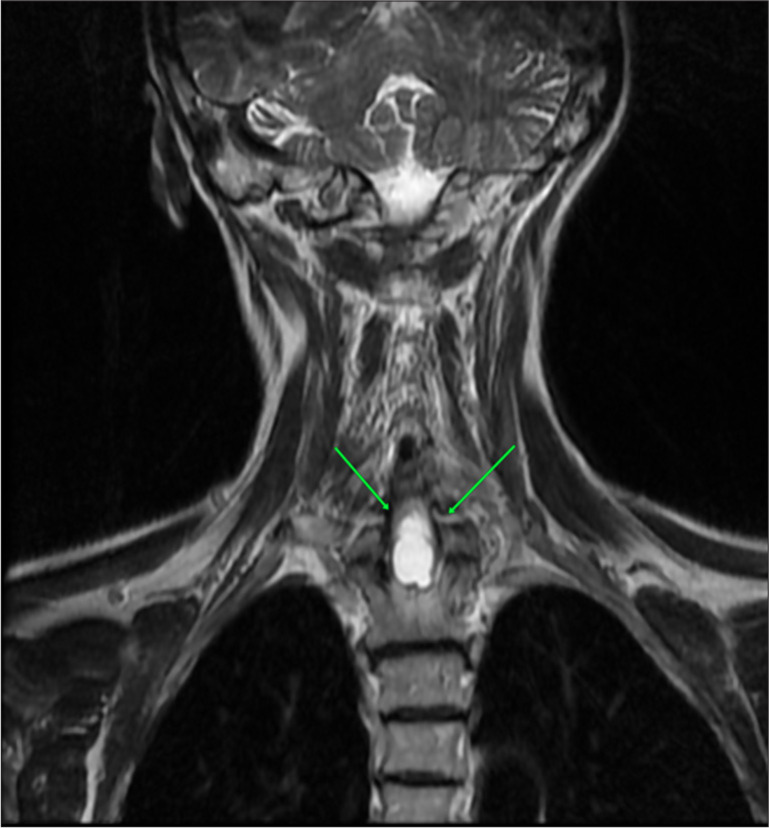
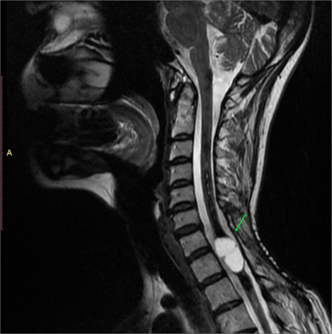
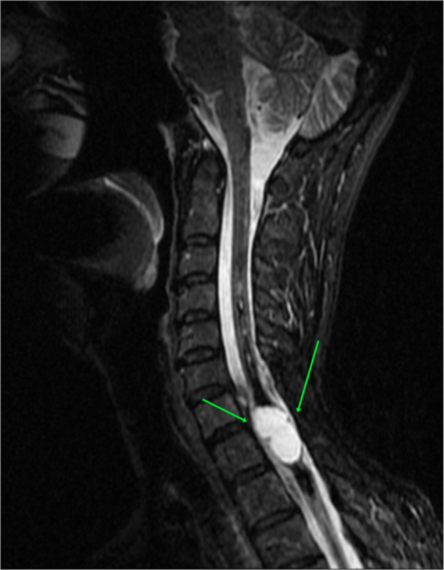
Syringomyelia is a rare condition characterized by the formation of a fluid-filled cyst within the spinal cord, leading to myelopathy. In addition, the pathological enlargement of the central canal is referred to as hydromyelia or cleft-like syrinx. We present a case of idiopathic syringomyelia and hematomyelia in a 50-year-old female patient with a 5-year follow-up on her disease progression. Magnetic resonance imaging (MRI) images revealed low-signal intensity on T1 and high-signal intensity on T2, with elevated hemorrhagic signal intensity on T1 and low peripheral signal intensity on T2. A fluid-filled lesion measuring 12 × 36 mm was observed between the C7 and Th3 vertebrae, with separation from some of the detailed components. No contrast enhancement was noted following IV contrast administration. Based on the MRI findings, a diagnosis consistent with giant hemorrhagic syringomyelia was established. Subsequently, a neurosurgical intervention was performed, resulting in a reduction in the size of the syringomyelia and a moderate improvement in the patient's symptom profile.
期刊介绍:
The Journal of Clinical Imaging Science (JCIS) is an open access peer-reviewed journal committed to publishing high-quality articles in the field of Imaging Science. The journal aims to present Imaging Science and relevant clinical information in an understandable and useful format. The journal is owned and published by the Scientific Scholar. Audience Our audience includes Radiologists, Researchers, Clinicians, medical professionals and students. Review process JCIS has a highly rigorous peer-review process that makes sure that manuscripts are scientifically accurate, relevant, novel and important. Authors disclose all conflicts, affiliations and financial associations such that the published content is not biased.

 求助内容:
求助内容: 应助结果提醒方式:


