Emika Murasawa, Komei Kameyama, Hajime Nakae, Naoko Mori
{"title":"Blunt traumatic inferior vena cava injury associated with seatbelt syndrome: The critical role of diagnosis and non-operative management.","authors":"Emika Murasawa, Komei Kameyama, Hajime Nakae, Naoko Mori","doi":"10.25259/JCIS_39_2025","DOIUrl":null,"url":null,"abstract":"<p><p>Blunt traumatic inferior vena cava (IVC) injury is rare and presents diagnostic and therapeutic challenges. We report a case of blunt traumatic IVC injury associated with bowel perforation and spinal cord injury, successfully managed with non-operative conservative treatment. A 57-year-old woman sustained injuries in a motor vehicle collision with a seatbelt fastened. Computed tomography (CT) revealed an irregular IVC contour at the infrarenal level and a retroperitoneal hematoma, leading to the diagnosis of blunt traumatic IVC injury. Free intraperitoneal air suggested bowel perforation, and magnetic resonance imaging confirmed a C5/6 spinal cord injury. This combination of injuries may raise suspicion for a seatbelt injury pattern. The bowel perforation was surgically treated, and posterior fixation was performed for the spinal injury. Since the patient remained hemodynamically stable, conservative management was selected for the IVC injury. Follow-up CT revealed a reduction in the retroperitoneal hematoma and improvement in the IVC contour, indicating successful conservative treatment. Blunt traumatic IVC injury is rare, and some cases do not exhibit contrast media extravasation. In this case, the diagnosis was based on IVC contour abnormalities and retroperitoneal hematoma. Considering the patient's stable hemodynamics, conservative treatment was selected. Careful interpretation of CT findings is essential for diagnosing IVC injury, and appropriate clinical judgment is key to achieving successful non-operative management in selected cases.</p>","PeriodicalId":15512,"journal":{"name":"Journal of Clinical Imaging Science","volume":"15 ","pages":"20"},"PeriodicalIF":1.3000,"publicationDate":"2025-06-02","publicationTypes":"Journal Article","fieldsOfStudy":null,"isOpenAccess":false,"openAccessPdf":"https://www.ncbi.nlm.nih.gov/pmc/articles/PMC12289102/pdf/","citationCount":"0","resultStr":null,"platform":"Semanticscholar","paperid":null,"PeriodicalName":"Journal of Clinical Imaging Science","FirstCategoryId":"1085","ListUrlMain":"https://doi.org/10.25259/JCIS_39_2025","RegionNum":0,"RegionCategory":null,"ArticlePicture":[],"TitleCN":null,"AbstractTextCN":null,"PMCID":null,"EPubDate":"2025/1/1 0:00:00","PubModel":"eCollection","JCR":"Q3","JCRName":"RADIOLOGY, NUCLEAR MEDICINE & MEDICAL IMAGING","Score":null,"Total":0}
引用次数: 0
Abstract
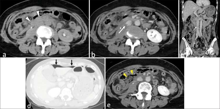
Blunt traumatic inferior vena cava (IVC) injury is rare and presents diagnostic and therapeutic challenges. We report a case of blunt traumatic IVC injury associated with bowel perforation and spinal cord injury, successfully managed with non-operative conservative treatment. A 57-year-old woman sustained injuries in a motor vehicle collision with a seatbelt fastened. Computed tomography (CT) revealed an irregular IVC contour at the infrarenal level and a retroperitoneal hematoma, leading to the diagnosis of blunt traumatic IVC injury. Free intraperitoneal air suggested bowel perforation, and magnetic resonance imaging confirmed a C5/6 spinal cord injury. This combination of injuries may raise suspicion for a seatbelt injury pattern. The bowel perforation was surgically treated, and posterior fixation was performed for the spinal injury. Since the patient remained hemodynamically stable, conservative management was selected for the IVC injury. Follow-up CT revealed a reduction in the retroperitoneal hematoma and improvement in the IVC contour, indicating successful conservative treatment. Blunt traumatic IVC injury is rare, and some cases do not exhibit contrast media extravasation. In this case, the diagnosis was based on IVC contour abnormalities and retroperitoneal hematoma. Considering the patient's stable hemodynamics, conservative treatment was selected. Careful interpretation of CT findings is essential for diagnosing IVC injury, and appropriate clinical judgment is key to achieving successful non-operative management in selected cases.
期刊介绍:
The Journal of Clinical Imaging Science (JCIS) is an open access peer-reviewed journal committed to publishing high-quality articles in the field of Imaging Science. The journal aims to present Imaging Science and relevant clinical information in an understandable and useful format. The journal is owned and published by the Scientific Scholar. Audience Our audience includes Radiologists, Researchers, Clinicians, medical professionals and students. Review process JCIS has a highly rigorous peer-review process that makes sure that manuscripts are scientifically accurate, relevant, novel and important. Authors disclose all conflicts, affiliations and financial associations such that the published content is not biased.


 求助内容:
求助内容: 应助结果提醒方式:


