The Integration of Micro-CT Imaging and Finite Element Simulations for Modelling Tooth-Inlay Systems for Mechanical Stress Analysis: A Preliminary Study.
Nikoleta Nikolova, Miryana Raykovska, Nikolay Petkov, Martin Tsvetkov, Ivan Georgiev, Eugeni Koytchev, Roumen Iankov, Mariana Dimova-Gabrovska, Angela Gusiyska
{"title":"The Integration of Micro-CT Imaging and Finite Element Simulations for Modelling Tooth-Inlay Systems for Mechanical Stress Analysis: A Preliminary Study.","authors":"Nikoleta Nikolova, Miryana Raykovska, Nikolay Petkov, Martin Tsvetkov, Ivan Georgiev, Eugeni Koytchev, Roumen Iankov, Mariana Dimova-Gabrovska, Angela Gusiyska","doi":"10.3390/jfb16070267","DOIUrl":null,"url":null,"abstract":"<p><p>This study presents a methodology for developing and validating digital models of tooth-inlay systems, aiming to trace the complete workflow from clinical procedures to simulation by involving dental professionals-dentists for manual cavity preparation and dental technicians for restoration modelling-while integrating micro-computed tomography (micro-CT) imaging with finite element analysis (FEA). The proposed workflow includes (1) the acquisition of high-resolution 3D micro-CT scans of a non-restored tooth, (2) image segmentation and reconstruction to create anatomically accurate digital twins and mesh generation, (3) the selection of proper resin and the 3D printing of four typodonts, (4) the manual preparation of cavities on the typodonts, (5) the acquisition of high-resolution 3D micro-CT scans of the typodonts, (6) mesh generation, digital inlay and onlay modelling and material property assignment, and (7) nonlinear FEA simulations under representative masticatory loading. The approach enables the visualisation of stress and deformation patterns, with preliminary results indicating stress concentrations at the tooth-restoration interface integrating different cavity alternatives and restorations on the same tooth. Quantitative outputs include von Mises stress, strain energy density, and displacement distribution. This study demonstrates the feasibility of using image-based, tooth-specific digital twins for biomechanical modelling in dentistry. The developed framework lays the groundwork for future investigations into the optimisation of restoration design and material selection in clinical applications.</p>","PeriodicalId":15767,"journal":{"name":"Journal of Functional Biomaterials","volume":"16 7","pages":""},"PeriodicalIF":5.2000,"publicationDate":"2025-07-21","publicationTypes":"Journal Article","fieldsOfStudy":null,"isOpenAccess":false,"openAccessPdf":"https://www.ncbi.nlm.nih.gov/pmc/articles/PMC12295181/pdf/","citationCount":"0","resultStr":null,"platform":"Semanticscholar","paperid":null,"PeriodicalName":"Journal of Functional Biomaterials","FirstCategoryId":"5","ListUrlMain":"https://doi.org/10.3390/jfb16070267","RegionNum":3,"RegionCategory":"医学","ArticlePicture":[],"TitleCN":null,"AbstractTextCN":null,"PMCID":null,"EPubDate":"","PubModel":"","JCR":"Q1","JCRName":"ENGINEERING, BIOMEDICAL","Score":null,"Total":0}
引用次数: 0
Abstract
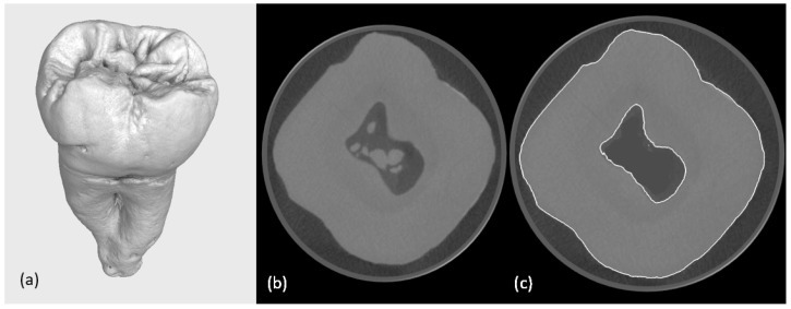
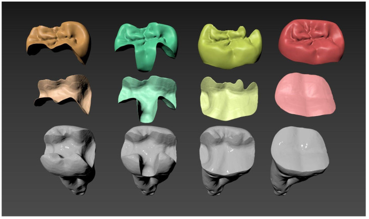
This study presents a methodology for developing and validating digital models of tooth-inlay systems, aiming to trace the complete workflow from clinical procedures to simulation by involving dental professionals-dentists for manual cavity preparation and dental technicians for restoration modelling-while integrating micro-computed tomography (micro-CT) imaging with finite element analysis (FEA). The proposed workflow includes (1) the acquisition of high-resolution 3D micro-CT scans of a non-restored tooth, (2) image segmentation and reconstruction to create anatomically accurate digital twins and mesh generation, (3) the selection of proper resin and the 3D printing of four typodonts, (4) the manual preparation of cavities on the typodonts, (5) the acquisition of high-resolution 3D micro-CT scans of the typodonts, (6) mesh generation, digital inlay and onlay modelling and material property assignment, and (7) nonlinear FEA simulations under representative masticatory loading. The approach enables the visualisation of stress and deformation patterns, with preliminary results indicating stress concentrations at the tooth-restoration interface integrating different cavity alternatives and restorations on the same tooth. Quantitative outputs include von Mises stress, strain energy density, and displacement distribution. This study demonstrates the feasibility of using image-based, tooth-specific digital twins for biomechanical modelling in dentistry. The developed framework lays the groundwork for future investigations into the optimisation of restoration design and material selection in clinical applications.
期刊介绍:
Journal of Functional Biomaterials (JFB, ISSN 2079-4983) is an international and interdisciplinary scientific journal that publishes regular research papers (articles), reviews and short communications about applications of materials for biomedical use. JFB covers subjects from chemistry, pharmacy, biology, physics over to engineering. The journal focuses on the preparation, performance and use of functional biomaterials in biomedical devices and their behaviour in physiological environments. Our aim is to encourage scientists to publish their results in as much detail as possible. Therefore, there is no restriction on the length of the papers. The full experimental details must be provided so that the results can be reproduced. Several topical special issues will be published. Scope: adhesion, adsorption, biocompatibility, biohybrid materials, bio-inert materials, biomaterials, biomedical devices, biomimetic materials, bone repair, cardiovascular devices, ceramics, composite materials, dental implants, dental materials, drug delivery systems, functional biopolymers, glasses, hyper branched polymers, molecularly imprinted polymers (MIPs), nanomedicine, nanoparticles, nanotechnology, natural materials, self-assembly smart materials, stimuli responsive materials, surface modification, tissue devices, tissue engineering, tissue-derived materials, urological devices.

 求助内容:
求助内容: 应助结果提醒方式:


