Peripheral TNF-α and CD8+/CD28+ T Lymphocytes as Alternatives for PD-L1 Prediction in Breast Cancer Tumor Microenvironment: Stratified by Neoadjuvant Therapy.
{"title":"Peripheral TNF-α and CD8<sup>+</sup>/CD28<sup>+</sup> T Lymphocytes as Alternatives for PD-L1 Prediction in Breast Cancer Tumor Microenvironment: Stratified by Neoadjuvant Therapy.","authors":"Jiangping Wu, Xin Ou, Keyu Yuan, Feng Shi, Quan Zhou, Suzhen Lyu, Yanping Li, Yanjie Zhao, Yu Cao, Jianping Sun, Qingkun Song","doi":"10.2147/BCTT.S532688","DOIUrl":null,"url":null,"abstract":"<p><strong>Background: </strong>Programmed death-ligand 1 (PD-L1) is an immunotherapy target; however, its detection is based on biopsy tissues, and repeated biopsies present clinical challenges. This study aimed to explore peripheral blood-based alternatives to PD-L1 tissue detection in breast cancer (BC), particularly stratification by neoadjuvant therapy (NAT).</p><p><strong>Methods: </strong>A total of 134 cases were recruited, the peripheral lymphocyte subtypes and cytokines were detected by flow cytometry and PD-L1 expression in tumor microenvironment (TME) was detected by immunohistochemistry and assessed by two qualified pathologists.</p><p><strong>Results: </strong>The patients with positive PD-L1 expression had peripheral CD8<sup>+</sup>/CD28<sup>+</sup> T lymphocytes 20% higher than those with negative expression (<i>p</i> = 0.008) with the area under the receiver operating characteristic curve (AUC) being 0.64 (<i>p</i> = 0.002). Among patients with negative NAT, positive PD-L1 expression was associated with peripheral CD8<sup>+</sup>/CD28<sup>+</sup> T lymphocytes that increased by 54% (<i>p</i> = 0.003), and the AUC being 0.68 (<i>p</i> = 0.003). In patients receiving NAT, positive PD-L1 expression was associated with peripheral TNF-α (<i>p</i> = 0.010), which increased from 0.45pg/mL to 0.64pg/mL in the PD-L1 positive group, and the AUC was 0.79 (<i>p</i> = 0.012). Among patients without NAT experience, a 1% increase in peripheral CD8<sup>+</sup>/CD28<sup>+</sup> T lymphocytes was associated with a 21% higher probability of positive PD-L1 expression (OR = 1.21, 95% CI: 1.06-1.37) and among patients with NAT, the OR of peripheral TNF-α (>0.5pg/mL) increased to 24.5 for positive TME PD-L1 expression (<i>p</i> = 0.008).</p><p><strong>Conclusion: </strong>Peripheral CD8<sup>+</sup>/CD28<sup>+</sup> T cell percentages and TNF-α levels served as non-invasive biomarkers for TME PD-L1 expression in BC patients with and without NAT, respectively. These biomarkers warranted further validation in clinical implementation to guide precision immunotherapy.</p>","PeriodicalId":9106,"journal":{"name":"Breast Cancer : Targets and Therapy","volume":"17 ","pages":"627-637"},"PeriodicalIF":3.4000,"publicationDate":"2025-07-19","publicationTypes":"Journal Article","fieldsOfStudy":null,"isOpenAccess":false,"openAccessPdf":"https://www.ncbi.nlm.nih.gov/pmc/articles/PMC12288228/pdf/","citationCount":"0","resultStr":null,"platform":"Semanticscholar","paperid":null,"PeriodicalName":"Breast Cancer : Targets and Therapy","FirstCategoryId":"3","ListUrlMain":"https://doi.org/10.2147/BCTT.S532688","RegionNum":4,"RegionCategory":"医学","ArticlePicture":[],"TitleCN":null,"AbstractTextCN":null,"PMCID":null,"EPubDate":"2025/1/1 0:00:00","PubModel":"eCollection","JCR":"Q2","JCRName":"ONCOLOGY","Score":null,"Total":0}
引用次数: 0
Abstract
Background: Programmed death-ligand 1 (PD-L1) is an immunotherapy target; however, its detection is based on biopsy tissues, and repeated biopsies present clinical challenges. This study aimed to explore peripheral blood-based alternatives to PD-L1 tissue detection in breast cancer (BC), particularly stratification by neoadjuvant therapy (NAT).
Methods: A total of 134 cases were recruited, the peripheral lymphocyte subtypes and cytokines were detected by flow cytometry and PD-L1 expression in tumor microenvironment (TME) was detected by immunohistochemistry and assessed by two qualified pathologists.
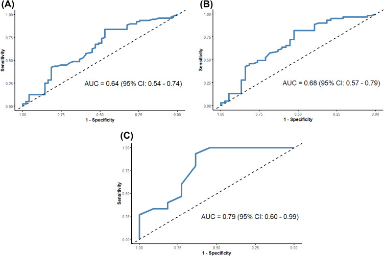
Results: The patients with positive PD-L1 expression had peripheral CD8+/CD28+ T lymphocytes 20% higher than those with negative expression (p = 0.008) with the area under the receiver operating characteristic curve (AUC) being 0.64 (p = 0.002). Among patients with negative NAT, positive PD-L1 expression was associated with peripheral CD8+/CD28+ T lymphocytes that increased by 54% (p = 0.003), and the AUC being 0.68 (p = 0.003). In patients receiving NAT, positive PD-L1 expression was associated with peripheral TNF-α (p = 0.010), which increased from 0.45pg/mL to 0.64pg/mL in the PD-L1 positive group, and the AUC was 0.79 (p = 0.012). Among patients without NAT experience, a 1% increase in peripheral CD8+/CD28+ T lymphocytes was associated with a 21% higher probability of positive PD-L1 expression (OR = 1.21, 95% CI: 1.06-1.37) and among patients with NAT, the OR of peripheral TNF-α (>0.5pg/mL) increased to 24.5 for positive TME PD-L1 expression (p = 0.008).
Conclusion: Peripheral CD8+/CD28+ T cell percentages and TNF-α levels served as non-invasive biomarkers for TME PD-L1 expression in BC patients with and without NAT, respectively. These biomarkers warranted further validation in clinical implementation to guide precision immunotherapy.

 求助内容:
求助内容: 应助结果提醒方式:


