Julia Pfirrmann, Anne Adlung, Ingo Hermann, Claudia E. Weber, Anne Ebert, Frank G. Zöllner, Achim Gass, Philipp Eisele
{"title":"Multimodal Quantitative MRI Atlas of the Human Brain in Healthy Young Adults","authors":"Julia Pfirrmann, Anne Adlung, Ingo Hermann, Claudia E. Weber, Anne Ebert, Frank G. Zöllner, Achim Gass, Philipp Eisele","doi":"10.1111/jon.70074","DOIUrl":null,"url":null,"abstract":"<div>\n \n \n <section>\n \n <h3> Background and Purpose</h3>\n \n <p>Quantitative MRI provides insights into (patho-) physiological processes of the brain macro- and microstructure. In this study, we applied a multimodal MRI approach to determine quantitative values of the brain in healthy young adults.</p>\n </section>\n \n <section>\n \n <h3> Methods</h3>\n \n <p>3 T MRI including sodium MRI (4.0 mm nominal isotropic resolution), 3D T1-weighted magnetization-prepared rapid acquisition gradient-echo (spatial resolution = 0.9 mm × 0.9 mm × 0.9 mm), T2-fluid attenuated inversion recovery (spatial resolution = 0.4 mm × 0.4 mm × 5.0 mm), diffusion-weighted imaging (spatial resolution = 1.0 mm × 1.0 mm × 4.0 mm), PD/T2 (spatial resolution = 1.0 mm × 1.0 mm × 2.0 mm), and MR fingerprinting including T1 and T2* relaxation times (spatial resolution = 1 mm × 1 mm × 2 mm) was performed on 40 healthy young adults (28 women, mean age: 24.55 years).</p>\n </section>\n \n <section>\n \n <h3> Results</h3>\n \n <p>Mean values for the gray matter were as follows—apparent diffusion coefficient (ADC): 0.95 ± 0.03 × 10<sup>−3</sup> mm<sup>2</sup>/s, total sodium concentration (TSC): 41.99 ± 2.35 mM, T1: 1338.04 ± 46.29 ms, T2*: 63.39 ± 2.94 ms. For the white matter, mean values were as follows—ADC: 0.79 ± 0.02 × 10<sup>−3</sup> mm<sup>2</sup>/s, TSC: 36.08 ± 5.62 mM, T1: 968.47 ± 48.35 ms, T2*: 53.62 ± 8.06 ms, and for the deep gray matter, mean values were as follows—ADC: 0.85 ± 0.04 × 10<sup>−3</sup> mm<sup>2</sup>/s, TSC: 38.23 ± 2.91 mM, T1: 1087.24 ± 39.77 ms, T2*: 54.53 ± 7.15.</p>\n </section>\n \n <section>\n \n <h3> Conclusion</h3>\n \n <p>Our multimodal, quantitative MRI atlas of the human brain in healthy young adults provides meaningful in vivo insights into the brain microstructure and can be used for reference in future studies.</p>\n </section>\n </div>","PeriodicalId":16399,"journal":{"name":"Journal of Neuroimaging","volume":"35 4","pages":""},"PeriodicalIF":2.3000,"publicationDate":"2025-07-25","publicationTypes":"Journal Article","fieldsOfStudy":null,"isOpenAccess":false,"openAccessPdf":"https://onlinelibrary.wiley.com/doi/epdf/10.1111/jon.70074","citationCount":"0","resultStr":null,"platform":"Semanticscholar","paperid":null,"PeriodicalName":"Journal of Neuroimaging","FirstCategoryId":"3","ListUrlMain":"https://onlinelibrary.wiley.com/doi/10.1111/jon.70074","RegionNum":4,"RegionCategory":"医学","ArticlePicture":[],"TitleCN":null,"AbstractTextCN":null,"PMCID":null,"EPubDate":"","PubModel":"","JCR":"Q3","JCRName":"CLINICAL NEUROLOGY","Score":null,"Total":0}
引用次数: 0
Abstract
Background and Purpose
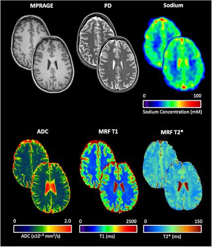
Quantitative MRI provides insights into (patho-) physiological processes of the brain macro- and microstructure. In this study, we applied a multimodal MRI approach to determine quantitative values of the brain in healthy young adults.
Methods
3 T MRI including sodium MRI (4.0 mm nominal isotropic resolution), 3D T1-weighted magnetization-prepared rapid acquisition gradient-echo (spatial resolution = 0.9 mm × 0.9 mm × 0.9 mm), T2-fluid attenuated inversion recovery (spatial resolution = 0.4 mm × 0.4 mm × 5.0 mm), diffusion-weighted imaging (spatial resolution = 1.0 mm × 1.0 mm × 4.0 mm), PD/T2 (spatial resolution = 1.0 mm × 1.0 mm × 2.0 mm), and MR fingerprinting including T1 and T2* relaxation times (spatial resolution = 1 mm × 1 mm × 2 mm) was performed on 40 healthy young adults (28 women, mean age: 24.55 years).
Results
Mean values for the gray matter were as follows—apparent diffusion coefficient (ADC): 0.95 ± 0.03 × 10⁻³ mm2/s, total sodium concentration (TSC): 41.99 ± 2.35 mM, T1: 1338.04 ± 46.29 ms, T2*: 63.39 ± 2.94 ms. For the white matter, mean values were as follows—ADC: 0.79 ± 0.02 × 10⁻³ mm2/s, TSC: 36.08 ± 5.62 mM, T1: 968.47 ± 48.35 ms, T2*: 53.62 ± 8.06 ms, and for the deep gray matter, mean values were as follows—ADC: 0.85 ± 0.04 × 10⁻³ mm2/s, TSC: 38.23 ± 2.91 mM, T1: 1087.24 ± 39.77 ms, T2*: 54.53 ± 7.15.
Conclusion
Our multimodal, quantitative MRI atlas of the human brain in healthy young adults provides meaningful in vivo insights into the brain microstructure and can be used for reference in future studies.
期刊介绍:
Start reading the Journal of Neuroimaging to learn the latest neurological imaging techniques. The peer-reviewed research is written in a practical clinical context, giving you the information you need on:
MRI
CT
Carotid Ultrasound and TCD
SPECT
PET
Endovascular Surgical Neuroradiology
Functional MRI
Xenon CT
and other new and upcoming neuroscientific modalities.The Journal of Neuroimaging addresses the full spectrum of human nervous system disease, including stroke, neoplasia, degenerating and demyelinating disease, epilepsy, tumors, lesions, infectious disease, cerebral vascular arterial diseases, toxic-metabolic disease, psychoses, dementias, heredo-familial disease, and trauma.Offering original research, review articles, case reports, neuroimaging CPCs, and evaluations of instruments and technology relevant to the nervous system, the Journal of Neuroimaging focuses on useful clinical developments and applications, tested techniques and interpretations, patient care, diagnostics, and therapeutics. Start reading today!

 求助内容:
求助内容: 应助结果提醒方式:


