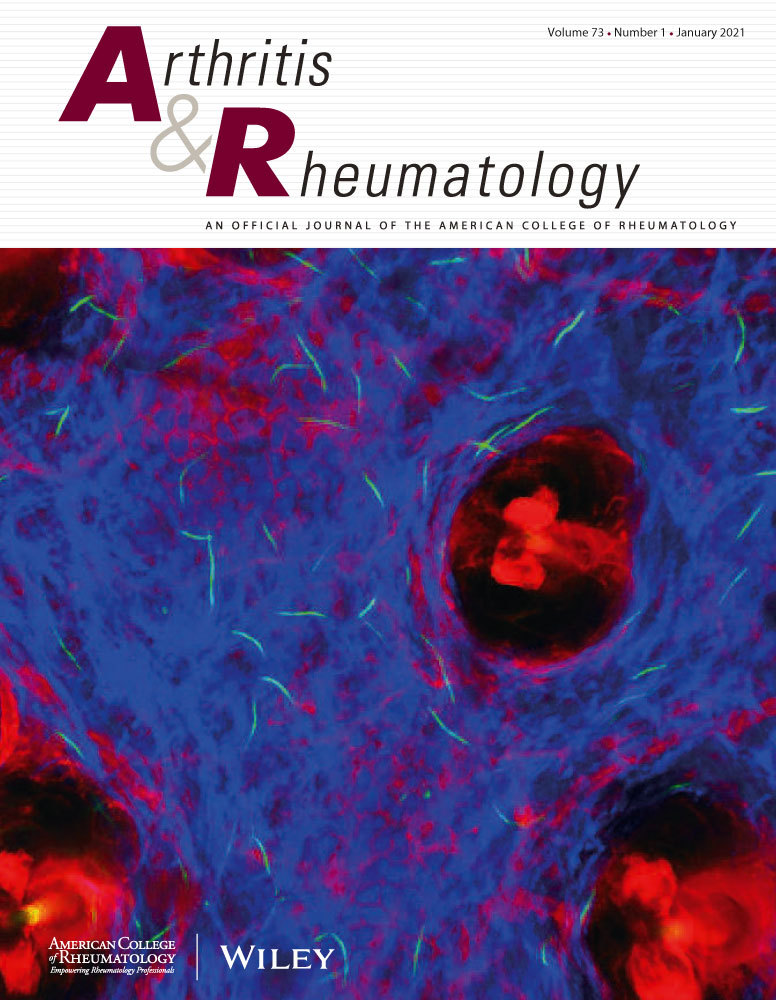
Serum urate levels alter the spatial distribution of urate crystals in synovium and correlate with synovitis and pain in non-gout female patients with anteromedial knee osteoarthritis.
IF 10.9
1区 医学
Q1 RHEUMATOLOGY
引用次数: 0
Abstract
OBJECTIVE This study aimed to investigate alterations in the spatial distribution of urate crystals in the osteoarthritic synovium of patients with different levels of serum uric acid (SUA). Additionally, we examined the association between SUA levels and severity of synovitis and pain in anteromedial osteoarthritis (AMOA) of the knee. METHODS Patients who underwent knee arthroplasty due to AMOA were prospectively enrolled. Blood, synovial fluid, and synovium samples were collected and UA levels were quantified. The degree of synovitis was evaluated histologically, and the levels of inflammatory cytokines in the synovial fluid were measured. The spatial distribution of urate crystals in the synovium was determined using Gomori methenamine silver staining, and the pain subscale of the Western Ontario McMaster Universities Osteoarthritis Index was used to evaluate knee pain. The relationships between SUA, degree of synovitis, and knee pain were assessed using Spearman rank correlation and multivariable analyses. RESULTS The pattern of urate crystal deposition in the osteoarthritic synovium was significantly altered in populations with different SUA levels. The capillary wall, sublining layer, and lining cells were sequentially affected. SUA level was the only risk factor for high-grade synovitis and severe pain in the multivariate analysis. SUA level was also positively correlated with UA level in the synovial fluid and synovium, Krenn histological score of the synovium, knee pain, and inflammatory cytokine level in the synovial fluid. CONCLUSION Spatial urate crystal distribution in the synovium was altered when SUA levels were elevated. Elevated SUA levels were also associated with aggravated synovitis and pain.血清尿酸水平改变了滑膜中尿酸盐晶体的空间分布,并与非痛风女性膝前内侧骨关节炎患者的滑膜炎和疼痛相关。
目的探讨不同血清尿酸(SUA)水平对骨关节炎滑膜尿酸晶体空间分布的影响。此外,我们研究了SUA水平与膝关节前内侧骨关节炎(AMOA)滑膜炎和疼痛严重程度之间的关系。方法前瞻性纳入因AMOA而行膝关节置换术的患者。采集血液、滑液和滑膜样本,定量测定UA水平。组织学上评估滑膜炎的程度,并测量滑膜液中炎症细胞因子的水平。采用Gomori甲基苯丙胺银染色法测定滑膜内尿酸盐晶体的空间分布,并采用Western Ontario McMaster university Osteoarthritis Index疼痛亚量表评估膝关节疼痛。采用Spearman秩相关和多变量分析评估SUA、滑膜炎程度和膝关节疼痛之间的关系。结果不同SUA水平人群骨关节炎滑膜中尿酸晶体沉积模式明显改变。毛细血管壁、下衬层和衬细胞依次受到影响。在多变量分析中,SUA水平是高度滑膜炎和剧烈疼痛的唯一危险因素。SUA水平也与滑膜液和滑膜中的UA水平、滑膜的Krenn组织学评分、膝关节疼痛和滑膜液中的炎症细胞因子水平呈正相关。结论当SUA水平升高时,滑膜内尿酸晶体的空间分布发生改变。SUA水平升高也与滑膜炎和疼痛加重有关。
本文章由计算机程序翻译,如有差异,请以英文原文为准。
求助全文
约1分钟内获得全文
求助全文
来源期刊

Arthritis & Rheumatology
RHEUMATOLOGY-
CiteScore
20.90
自引率
3.00%
发文量
371
期刊介绍:
Arthritis & Rheumatology is the official journal of the American College of Rheumatology and focuses on the natural history, pathophysiology, treatment, and outcome of rheumatic diseases. It is a peer-reviewed publication that aims to provide the highest quality basic and clinical research in this field. The journal covers a wide range of investigative areas and also includes review articles, editorials, and educational material for researchers and clinicians. Being recognized as a leading research journal in rheumatology, Arthritis & Rheumatology serves the global community of rheumatology investigators and clinicians.
 求助内容:
求助内容: 应助结果提醒方式:


