Yumeng Yan, Roberto Rotundo, Jeanie Suvan, Marco Orlandi, Alessandro Poma, Francesco D'Aiuto
{"title":"Photodynamic therapy and peri-implant diseases: a systematic review and meta-analysis.","authors":"Yumeng Yan, Roberto Rotundo, Jeanie Suvan, Marco Orlandi, Alessandro Poma, Francesco D'Aiuto","doi":"10.3389/froh.2025.1614982","DOIUrl":null,"url":null,"abstract":"<p><strong>Aim: </strong>This systematic review aimed to evaluate the antimicrobial efficacy of photodynamic therapy (PDT) in treating peri-implant diseases when combined with mechanical debridement (MD) compared with mechanical debridement alone.</p><p><strong>Methods: </strong>A systematic review was completed according to PRISMA guidelines. The following databases, Cochrane Central Register for Controlled Trials (CENTRAL), Medline, Embase, Dentistry & Oral Sciences Source, Scopus, LILACS, and China Online, were searched based on the search strategies and hand search without language limitation until 15 June 2024. Only randomised controlled trials were included, assessing the efficacy of PDT used in combination with either surgical or non-surgical MD, compared with MD alone in participants with peri-implant diseases. Risk of bias for randomised controlled trials was assessed according to the recommendation of the Cochrane Reviewers' Handbook using the revised Cochrane tool. All outcomes were evaluated using the Grading of Recommendations Assessment, Development, and Evaluation (GRADE) approach.</p><p><strong>Results: </strong>A total of 26 studies were included in this study, of which 6 were categorised as low risk of bias, 9 were with some concern, and 11 were at high risk of bias. Nineteen studies were included in the quantitative analysis. At 3 months, PDT combined with non-surgical MD significantly reduced probing pocket depth (PPD) in peri-implant mucositis (-0.95 mm, 95% CI: -1.76 to -0.14) and peri-implantitis (-0.86 mm, 95% CI: -1.21 to -0.51) compared with MD alone. At 6 months, PPD reductions in peri-implantitis remained significant with non-surgical MD + PDT (-0.83 mm, 95% CI: -1.62 to -0.04) and surgical MD + PDT (-0.56 mm, 95% CI: -0.85 to -0.27). Non-surgical MD + PDT also reduced bleeding on probing (BoP) (-11.65% at 3 months, -6.76% at 6 months) and crestal bone loss (CBL) (-0.24 mm at 3 months, -0.28 mm at 6 months).</p><p><strong>Conclusion: </strong>PDT enhances antimicrobial efficacy in peri-implant disease treatment, significantly improving PPD, CBL, and BoP when combined with MD. However, due to the overall moderate-to-low certainty of the evidence and some concerns regarding risk of bias in the included studies, these findings should be interpreted with caution. Further high-quality, well-designed randomised controlled trials are warranted to confirm these effects and optimise treatment protocols.</p><p><strong>Systematic review registration: </strong>PROSPERO CRD42021262889.</p>","PeriodicalId":94016,"journal":{"name":"Frontiers in oral health","volume":"6 ","pages":"1614982"},"PeriodicalIF":3.1000,"publicationDate":"2025-07-09","publicationTypes":"Journal Article","fieldsOfStudy":null,"isOpenAccess":false,"openAccessPdf":"https://www.ncbi.nlm.nih.gov/pmc/articles/PMC12283991/pdf/","citationCount":"0","resultStr":null,"platform":"Semanticscholar","paperid":null,"PeriodicalName":"Frontiers in oral health","FirstCategoryId":"1085","ListUrlMain":"https://doi.org/10.3389/froh.2025.1614982","RegionNum":0,"RegionCategory":null,"ArticlePicture":[],"TitleCN":null,"AbstractTextCN":null,"PMCID":null,"EPubDate":"2025/1/1 0:00:00","PubModel":"eCollection","JCR":"Q1","JCRName":"DENTISTRY, ORAL SURGERY & MEDICINE","Score":null,"Total":0}
引用次数: 0
Abstract
Aim: This systematic review aimed to evaluate the antimicrobial efficacy of photodynamic therapy (PDT) in treating peri-implant diseases when combined with mechanical debridement (MD) compared with mechanical debridement alone.
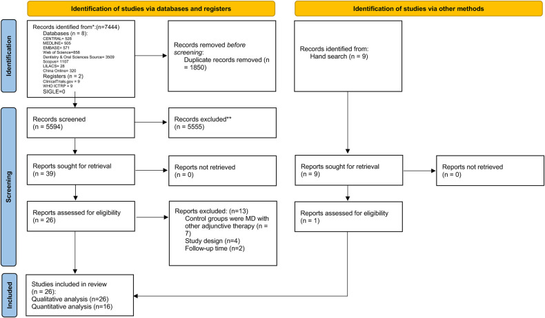
Methods: A systematic review was completed according to PRISMA guidelines. The following databases, Cochrane Central Register for Controlled Trials (CENTRAL), Medline, Embase, Dentistry & Oral Sciences Source, Scopus, LILACS, and China Online, were searched based on the search strategies and hand search without language limitation until 15 June 2024. Only randomised controlled trials were included, assessing the efficacy of PDT used in combination with either surgical or non-surgical MD, compared with MD alone in participants with peri-implant diseases. Risk of bias for randomised controlled trials was assessed according to the recommendation of the Cochrane Reviewers' Handbook using the revised Cochrane tool. All outcomes were evaluated using the Grading of Recommendations Assessment, Development, and Evaluation (GRADE) approach.
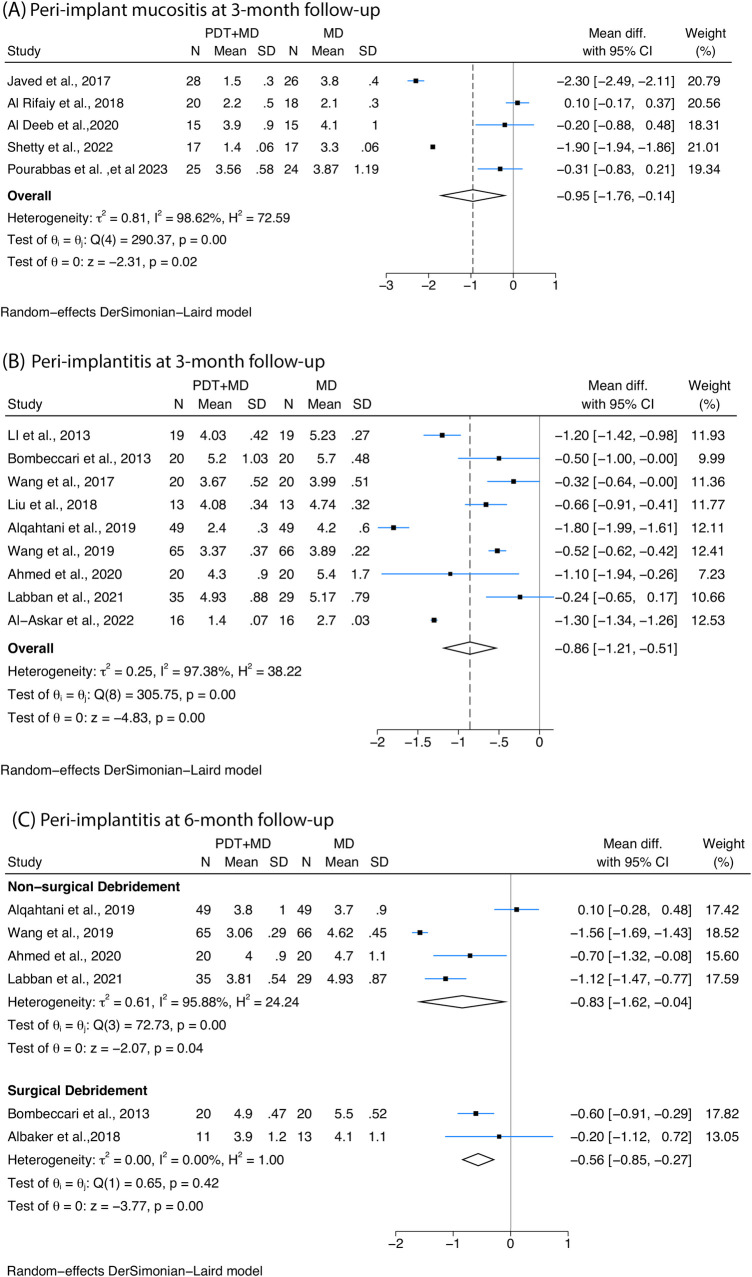
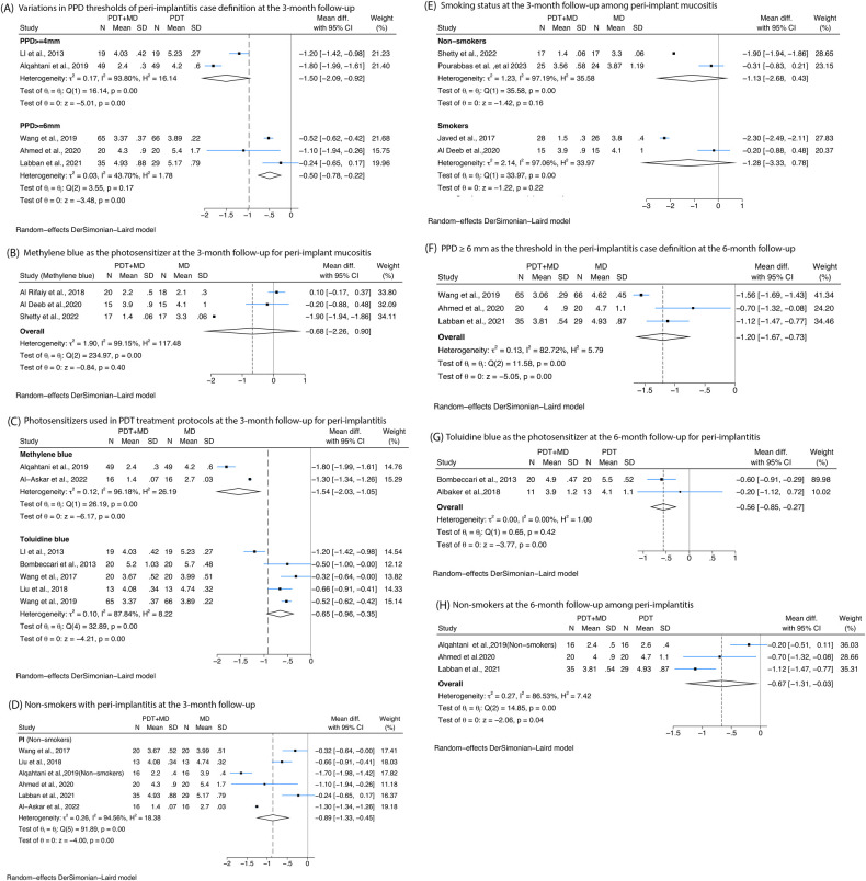
Results: A total of 26 studies were included in this study, of which 6 were categorised as low risk of bias, 9 were with some concern, and 11 were at high risk of bias. Nineteen studies were included in the quantitative analysis. At 3 months, PDT combined with non-surgical MD significantly reduced probing pocket depth (PPD) in peri-implant mucositis (-0.95 mm, 95% CI: -1.76 to -0.14) and peri-implantitis (-0.86 mm, 95% CI: -1.21 to -0.51) compared with MD alone. At 6 months, PPD reductions in peri-implantitis remained significant with non-surgical MD + PDT (-0.83 mm, 95% CI: -1.62 to -0.04) and surgical MD + PDT (-0.56 mm, 95% CI: -0.85 to -0.27). Non-surgical MD + PDT also reduced bleeding on probing (BoP) (-11.65% at 3 months, -6.76% at 6 months) and crestal bone loss (CBL) (-0.24 mm at 3 months, -0.28 mm at 6 months).
Conclusion: PDT enhances antimicrobial efficacy in peri-implant disease treatment, significantly improving PPD, CBL, and BoP when combined with MD. However, due to the overall moderate-to-low certainty of the evidence and some concerns regarding risk of bias in the included studies, these findings should be interpreted with caution. Further high-quality, well-designed randomised controlled trials are warranted to confirm these effects and optimise treatment protocols.

 求助内容:
求助内容: 应助结果提醒方式:


