{"title":"Effect of two deproteinized bone graft materials for socket preservation: a clinical and histological study.","authors":"Marwa Madi, Nasser S Al-Naief, Adel S Alagl","doi":"10.3389/froh.2025.1630504","DOIUrl":null,"url":null,"abstract":"<p><strong>Objective: </strong>To evaluate dimensional changes and new bone formation using two deproteinized bovine bone minerals, NuOss and Bio-Oss, in socket preservation.</p><p><strong>Materials and methods: </strong>Eighteen patients (6 males, 12 females; aged 23-45 years) requiring posterior tooth extraction were enrolled. Eighteen extraction sockets were augmented with either NuOss or Bio-Oss and covered with a collagen membrane. After six months, Cone Beam Cephalometry (CBCT) assessed dimensional changes in buccolingual width and buccal bone thickness. Bone core biopsies were obtained during implant placement and decalcified for histomorphologic examination. Statistical analysis compared dimensional changes and histomorphometric parameters between groups.</p><p><strong>Results: </strong>All experimental sites healed uneventfully, with complete soft tissue healing within four weeks and successful implant placement. CBCT scans showed comparable, non-significant dimensional reductions. Histomorphologic examination revealed lamellar cortical bone and osteoid trabeculae with partial to optimal integration. NuOss demonstrated significantly higher new bone formation (52.5 ± 2.5%) compared to Bio-Oss (37.5 ± 2.5%; <i>p</i> = 0.0021), with lower residual graft material (27.5 ± 2.5% vs. 42.5 ± 2.5%; <i>p</i> = 0.0018). Bio-Oss grafted cases exhibited more pronounced inflammatory cell infiltration. Soft tissue proportions were similar between groups (NuOss: 22.5 ± 2.5%, Bio-Oss: 17.5 ± 2.5%; <i>p</i> = 0.0892).</p><p><strong>Conclusion: </strong>Both NuOss and Bio-Oss showed positive bone regeneration effects. However, NuOss demonstrated more favorable biocompatibility, with less inflammation and improved bone integration than Bio-Oss.</p>","PeriodicalId":94016,"journal":{"name":"Frontiers in oral health","volume":"6 ","pages":"1630504"},"PeriodicalIF":3.1000,"publicationDate":"2025-07-09","publicationTypes":"Journal Article","fieldsOfStudy":null,"isOpenAccess":false,"openAccessPdf":"https://www.ncbi.nlm.nih.gov/pmc/articles/PMC12283978/pdf/","citationCount":"0","resultStr":null,"platform":"Semanticscholar","paperid":null,"PeriodicalName":"Frontiers in oral health","FirstCategoryId":"1085","ListUrlMain":"https://doi.org/10.3389/froh.2025.1630504","RegionNum":0,"RegionCategory":null,"ArticlePicture":[],"TitleCN":null,"AbstractTextCN":null,"PMCID":null,"EPubDate":"2025/1/1 0:00:00","PubModel":"eCollection","JCR":"Q1","JCRName":"DENTISTRY, ORAL SURGERY & MEDICINE","Score":null,"Total":0}
引用次数: 0
Abstract
Objective: To evaluate dimensional changes and new bone formation using two deproteinized bovine bone minerals, NuOss and Bio-Oss, in socket preservation.
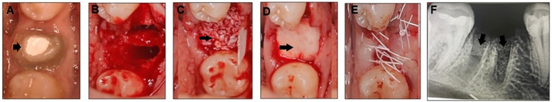
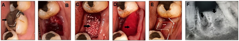
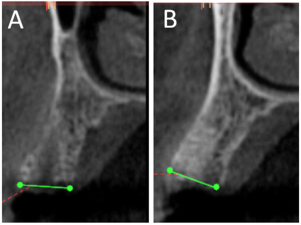
Materials and methods: Eighteen patients (6 males, 12 females; aged 23-45 years) requiring posterior tooth extraction were enrolled. Eighteen extraction sockets were augmented with either NuOss or Bio-Oss and covered with a collagen membrane. After six months, Cone Beam Cephalometry (CBCT) assessed dimensional changes in buccolingual width and buccal bone thickness. Bone core biopsies were obtained during implant placement and decalcified for histomorphologic examination. Statistical analysis compared dimensional changes and histomorphometric parameters between groups.
Results: All experimental sites healed uneventfully, with complete soft tissue healing within four weeks and successful implant placement. CBCT scans showed comparable, non-significant dimensional reductions. Histomorphologic examination revealed lamellar cortical bone and osteoid trabeculae with partial to optimal integration. NuOss demonstrated significantly higher new bone formation (52.5 ± 2.5%) compared to Bio-Oss (37.5 ± 2.5%; p = 0.0021), with lower residual graft material (27.5 ± 2.5% vs. 42.5 ± 2.5%; p = 0.0018). Bio-Oss grafted cases exhibited more pronounced inflammatory cell infiltration. Soft tissue proportions were similar between groups (NuOss: 22.5 ± 2.5%, Bio-Oss: 17.5 ± 2.5%; p = 0.0892).
Conclusion: Both NuOss and Bio-Oss showed positive bone regeneration effects. However, NuOss demonstrated more favorable biocompatibility, with less inflammation and improved bone integration than Bio-Oss.

 求助内容:
求助内容: 应助结果提醒方式:


