Extremely Rare Case of a Giant Paratubal Cyst, Coexisting with a Mucinous Cystadenoma, Surgically Treated Through Laparoscopy-A Case Report and Review of the Literature.
{"title":"Extremely Rare Case of a Giant Paratubal Cyst, Coexisting with a Mucinous Cystadenoma, Surgically Treated Through Laparoscopy-A Case Report and Review of the Literature.","authors":"Tudor Andrei Butureanu, Ana-Maria Apetrei, Ioana Pavaleanu, Ana-Maria Haliciu, Razvan Socolov, Raluca Balan","doi":"10.3390/reports8030106","DOIUrl":null,"url":null,"abstract":"<p><p><b>Background and Clinical Significance</b>: A paratubal cyst, which makes up about 10% of all adnexal masses, is a specific type of adnexal cyst that develops from the mesothelium in the broad ligament located between the fallopian tube and the ovary. Interestingly, the majority of paratubal cyst cases are initially misidentified as ovarian cysts, with suspicion arising in only 1 out of every 15 patients before undergoing surgery. <b>Case Presentation</b>: We report a case of a giant paratubal cyst mimicking an ovarian cyst in a 21-year-old woman supported by some representative images along with a literature review. The cyst's therapeutic management was surgical removal of the adnexa and the final postoperative histopathological diagnosis was that of a benign paratubal cyst. <b>Conclusions</b>: This case highlights the need to include a paratubal cyst in the differential diagnosis of pelvic masses, especially in women of reproductive age. To the best of our knowledge, this represents the largest paratubal cyst reported in the literature to date, based on overall dimensions and the highest recorded volume of aspirated fluid, successfully managed via laparoscopy. A further notable aspect of this case is the coexistence of the giant paratubal cyst with an ovarian mucinous cystadenoma.</p>","PeriodicalId":74664,"journal":{"name":"Reports (MDPI)","volume":"8 3","pages":""},"PeriodicalIF":0.8000,"publicationDate":"2025-07-14","publicationTypes":"Journal Article","fieldsOfStudy":null,"isOpenAccess":false,"openAccessPdf":"https://www.ncbi.nlm.nih.gov/pmc/articles/PMC12265995/pdf/","citationCount":"0","resultStr":null,"platform":"Semanticscholar","paperid":null,"PeriodicalName":"Reports (MDPI)","FirstCategoryId":"1085","ListUrlMain":"https://doi.org/10.3390/reports8030106","RegionNum":0,"RegionCategory":null,"ArticlePicture":[],"TitleCN":null,"AbstractTextCN":null,"PMCID":null,"EPubDate":"","PubModel":"","JCR":"Q3","JCRName":"MEDICINE, GENERAL & INTERNAL","Score":null,"Total":0}
引用次数: 0
Abstract
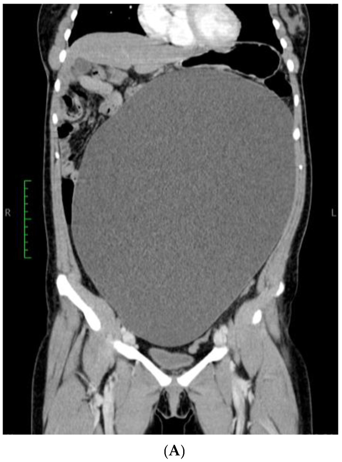
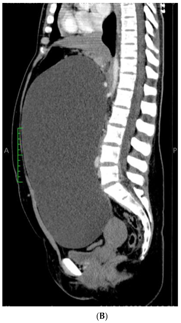
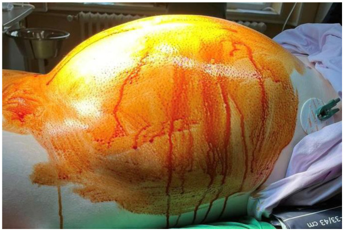
Background and Clinical Significance: A paratubal cyst, which makes up about 10% of all adnexal masses, is a specific type of adnexal cyst that develops from the mesothelium in the broad ligament located between the fallopian tube and the ovary. Interestingly, the majority of paratubal cyst cases are initially misidentified as ovarian cysts, with suspicion arising in only 1 out of every 15 patients before undergoing surgery. Case Presentation: We report a case of a giant paratubal cyst mimicking an ovarian cyst in a 21-year-old woman supported by some representative images along with a literature review. The cyst's therapeutic management was surgical removal of the adnexa and the final postoperative histopathological diagnosis was that of a benign paratubal cyst. Conclusions: This case highlights the need to include a paratubal cyst in the differential diagnosis of pelvic masses, especially in women of reproductive age. To the best of our knowledge, this represents the largest paratubal cyst reported in the literature to date, based on overall dimensions and the highest recorded volume of aspirated fluid, successfully managed via laparoscopy. A further notable aspect of this case is the coexistence of the giant paratubal cyst with an ovarian mucinous cystadenoma.

 求助内容:
求助内容: 应助结果提醒方式:


