Olli A Rantula, Jukka A Lipponen, Jari Halonen, Helena Jäntti, Tuomas T Rissanen, Noora S Naukkarinen, Eemu-Samuli Väliaho, Onni E Santala, Jagdeep Sedha, Tero J Martikainen, Juha E K Hartikainen
{"title":"Photoplethysmography in recent-onset atrial fibrillation: automatic detection of rhythm change and burden.","authors":"Olli A Rantula, Jukka A Lipponen, Jari Halonen, Helena Jäntti, Tuomas T Rissanen, Noora S Naukkarinen, Eemu-Samuli Väliaho, Onni E Santala, Jagdeep Sedha, Tero J Martikainen, Juha E K Hartikainen","doi":"10.1093/ehjdh/ztaf055","DOIUrl":null,"url":null,"abstract":"<p><strong>Aims: </strong>Atrial fibrillation (AF) is the most common arrhythmia, increasing stroke risk. Detecting AF is challenging due to its asymptomatic and paroxysmal nature. This study combines photoplethysmography (PPG) with automated techniques to detect AF, assess AF burden, and monitor rhythm changes from AF to sinus rhythm (SR).</p><p><strong>Methods and results: </strong>Ninety patients with recent-onset (duration <48 h) AF, scheduled for cardioversion, were monitored using a three-channel PPG armband on the upper arm. An ambulatory three-lead electrocardiogram (ECG) served as the gold standard. PPG recordings were segmented into 10-, 20-, 30-, and 60-min detection windows. Automated detection identified SR and AF episodes, rhythm changes, and AF burden. Sensitivities, specificities, positive predictive values (PPVs), and negative predictive values (NPVs) for rhythm detection were calculated, and the intraclass correlation coefficients (ICCs) for PPG-based AF burden were compared to the gold standard. Monitoring time ranged from 1.0 to 8.2 h per patient. Sensitivities, specificities, PPVs, and NPVs for AF detection were 93.9-94.6, 99.5-99.8, 99.4-99.7, and 93.7-95.0%, respectively. The ICC (0.97-0.98) indicated excellent agreement between PPG and the gold standard in estimating AF burden, with differences of -6.3 to -8.3 min (5.5-6.8%). Rhythm changes from AF to SR were detected in all patients (sensitivity 100%), with detection delays of 4.1 ± 1.4, 8.7 ± 2.8, 13.7 ± 3.9, and 27.8 ± 7.1 min depending on the detection window.</p><p><strong>Conclusion: </strong>Photoplethysmography with automated analysis shows promise in detecting AF, AF burden, and rhythm changes, indicating its potential in AF screening.</p><p><strong>Clinical trial registration: </strong>NCT04917653.</p>","PeriodicalId":72965,"journal":{"name":"European heart journal. Digital health","volume":"6 4","pages":"723-732"},"PeriodicalIF":4.4000,"publicationDate":"2025-05-23","publicationTypes":"Journal Article","fieldsOfStudy":null,"isOpenAccess":false,"openAccessPdf":"https://www.ncbi.nlm.nih.gov/pmc/articles/PMC12282364/pdf/","citationCount":"0","resultStr":null,"platform":"Semanticscholar","paperid":null,"PeriodicalName":"European heart journal. Digital health","FirstCategoryId":"1085","ListUrlMain":"https://doi.org/10.1093/ehjdh/ztaf055","RegionNum":0,"RegionCategory":null,"ArticlePicture":[],"TitleCN":null,"AbstractTextCN":null,"PMCID":null,"EPubDate":"2025/7/1 0:00:00","PubModel":"eCollection","JCR":"Q1","JCRName":"CARDIAC & CARDIOVASCULAR SYSTEMS","Score":null,"Total":0}
引用次数: 0
Abstract
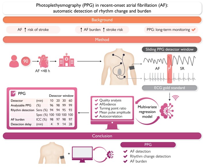
Aims: Atrial fibrillation (AF) is the most common arrhythmia, increasing stroke risk. Detecting AF is challenging due to its asymptomatic and paroxysmal nature. This study combines photoplethysmography (PPG) with automated techniques to detect AF, assess AF burden, and monitor rhythm changes from AF to sinus rhythm (SR).
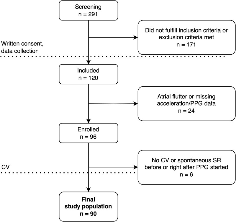
Methods and results: Ninety patients with recent-onset (duration <48 h) AF, scheduled for cardioversion, were monitored using a three-channel PPG armband on the upper arm. An ambulatory three-lead electrocardiogram (ECG) served as the gold standard. PPG recordings were segmented into 10-, 20-, 30-, and 60-min detection windows. Automated detection identified SR and AF episodes, rhythm changes, and AF burden. Sensitivities, specificities, positive predictive values (PPVs), and negative predictive values (NPVs) for rhythm detection were calculated, and the intraclass correlation coefficients (ICCs) for PPG-based AF burden were compared to the gold standard. Monitoring time ranged from 1.0 to 8.2 h per patient. Sensitivities, specificities, PPVs, and NPVs for AF detection were 93.9-94.6, 99.5-99.8, 99.4-99.7, and 93.7-95.0%, respectively. The ICC (0.97-0.98) indicated excellent agreement between PPG and the gold standard in estimating AF burden, with differences of -6.3 to -8.3 min (5.5-6.8%). Rhythm changes from AF to SR were detected in all patients (sensitivity 100%), with detection delays of 4.1 ± 1.4, 8.7 ± 2.8, 13.7 ± 3.9, and 27.8 ± 7.1 min depending on the detection window.
Conclusion: Photoplethysmography with automated analysis shows promise in detecting AF, AF burden, and rhythm changes, indicating its potential in AF screening.

 求助内容:
求助内容: 应助结果提醒方式:


