Butylated Hydroxyanisole (BHA) Disrupts Brain Signalling in Embryo-Larval Stage of Zebrafish Leading to Attention Deficit Hyperactivity Disorder (ADHD).
{"title":"Butylated Hydroxyanisole (BHA) Disrupts Brain Signalling in Embryo-Larval Stage of Zebrafish Leading to Attention Deficit Hyperactivity Disorder (ADHD).","authors":"Kandhasamy Veshaal, Ramasamy Vasantharekha, Usha Rani Balu, Mahesh Vallabi Aayush, Saheshnu Sai Balaji Pillai, Winkins Santosh, Barathi Seetharaman","doi":"10.3390/jox15040116","DOIUrl":null,"url":null,"abstract":"<p><strong>Background: </strong>Butylated hydroxyanisole (BHA) has been extensively used in several commercial industries as a preservative. It causes severe cellular and neurological damage affecting the developing fetus and might induce attention deficit hyperactivity disorder (ADHD).</p><p><strong>Methods: </strong>Zebrafish embryos were subjected to five distinct doses of BHA-0.5, 1, 2, 4, and 8 ppb up to 96 h post fertilization (hpf). Hatching rate, heart rate, and body malformations were assessed at 48 hpf, 72 hpf, and 48-96 hpf, respectively. After exposure, apoptotic activity, neurobehavioral evaluation, neurotransmitter assay, and antioxidant activity were assessed at 96 hpf. At 120 hpf, the expression of genes DRD4, COMT, 5-HTR1aa, and BDNF was evaluated by real-time PCR.</p><p><strong>Results: </strong>BHA exposure showed a delay in the hatching rate and a decrease in the heart rate of the embryo when compared with the control. Larvae exhibited developmental deformities such as bent spine, yolk sac, and pericardial edema. A higher density of apoptotic cells was observed in BHA-exposed larvae at 96 hpf. There was a decline in catalase (CAT), glutathione peroxidase (GPx), glutathione-S-transferase (GST), and superoxide dismutase (SOD) activity, indicating oxidative stress. There was a significant decrease in Acetylcholinesterase (AChE) activity and serotonin levels with an increase in concentration of BHA, leading to a dose-responsive increase in anxiety and impairment in memory. A significant decrease in gene expression was also observed for DRD4, COMT, 5-HTR1aa, and BDNF.</p><p><strong>Conclusions: </strong>Even at lower concentrations of BHA, zebrafish embryos suffered from developmental toxicity, anxiety, and impaired memory due to a decrease in AChE activity and serotonin levels and altered the expression of the mentioned genes.</p>","PeriodicalId":42356,"journal":{"name":"Journal of Xenobiotics","volume":"15 4","pages":""},"PeriodicalIF":4.4000,"publicationDate":"2025-07-09","publicationTypes":"Journal Article","fieldsOfStudy":null,"isOpenAccess":false,"openAccessPdf":"https://www.ncbi.nlm.nih.gov/pmc/articles/PMC12286280/pdf/","citationCount":"0","resultStr":null,"platform":"Semanticscholar","paperid":null,"PeriodicalName":"Journal of Xenobiotics","FirstCategoryId":"1085","ListUrlMain":"https://doi.org/10.3390/jox15040116","RegionNum":0,"RegionCategory":null,"ArticlePicture":[],"TitleCN":null,"AbstractTextCN":null,"PMCID":null,"EPubDate":"","PubModel":"","JCR":"Q1","JCRName":"TOXICOLOGY","Score":null,"Total":0}
引用次数: 0
Abstract
Background: Butylated hydroxyanisole (BHA) has been extensively used in several commercial industries as a preservative. It causes severe cellular and neurological damage affecting the developing fetus and might induce attention deficit hyperactivity disorder (ADHD).
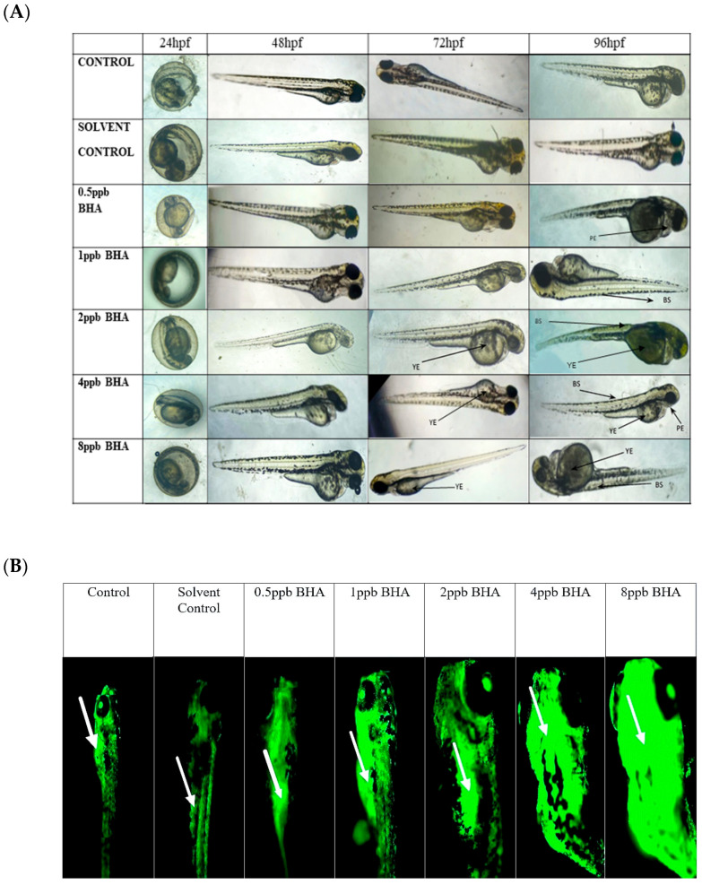
Methods: Zebrafish embryos were subjected to five distinct doses of BHA-0.5, 1, 2, 4, and 8 ppb up to 96 h post fertilization (hpf). Hatching rate, heart rate, and body malformations were assessed at 48 hpf, 72 hpf, and 48-96 hpf, respectively. After exposure, apoptotic activity, neurobehavioral evaluation, neurotransmitter assay, and antioxidant activity were assessed at 96 hpf. At 120 hpf, the expression of genes DRD4, COMT, 5-HTR1aa, and BDNF was evaluated by real-time PCR.
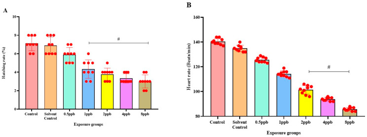
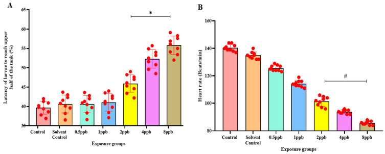
Results: BHA exposure showed a delay in the hatching rate and a decrease in the heart rate of the embryo when compared with the control. Larvae exhibited developmental deformities such as bent spine, yolk sac, and pericardial edema. A higher density of apoptotic cells was observed in BHA-exposed larvae at 96 hpf. There was a decline in catalase (CAT), glutathione peroxidase (GPx), glutathione-S-transferase (GST), and superoxide dismutase (SOD) activity, indicating oxidative stress. There was a significant decrease in Acetylcholinesterase (AChE) activity and serotonin levels with an increase in concentration of BHA, leading to a dose-responsive increase in anxiety and impairment in memory. A significant decrease in gene expression was also observed for DRD4, COMT, 5-HTR1aa, and BDNF.
Conclusions: Even at lower concentrations of BHA, zebrafish embryos suffered from developmental toxicity, anxiety, and impaired memory due to a decrease in AChE activity and serotonin levels and altered the expression of the mentioned genes.
期刊介绍:
The Journal of Xenobiotics publishes original studies concerning the beneficial (pharmacology) and detrimental effects (toxicology) of xenobiotics in all organisms. A xenobiotic (“stranger to life”) is defined as a chemical that is not usually found at significant concentrations or expected to reside for long periods in organisms. In addition to man-made chemicals, natural products could also be of interest if they have potent biological properties, special medicinal properties or that a given organism is at risk of exposure in the environment. Topics dealing with abiotic- and biotic-based transformations in various media (xenobiochemistry) and environmental toxicology are also of interest. Areas of interests include the identification of key physical and chemical properties of molecules that predict biological effects and persistence in the environment; the molecular mode of action of xenobiotics; biochemical and physiological interactions leading to change in organism health; pathophysiological interactions of natural and synthetic chemicals; development of biochemical indicators including new “-omics” approaches to identify biomarkers of exposure or effects for xenobiotics.

 求助内容:
求助内容: 应助结果提醒方式:


