Caterina Damiani, Cesare Ariasi, Giuseppe La Rosa, Francesca Di Lauro, Mariachiara Arisi, Vincenzo Maione, Marina Venturini, Simone Soglia
{"title":"Non-Invasive Diagnostic Techniques in Penile Intraepithelial Neoplasia (PeIN): Insights from Reflectance Confocal Microscopy (RCM), Line-Field Confocal Optical Coherence Tomography (LC-OCT), and Correlation with Histopathological Features.","authors":"Caterina Damiani, Cesare Ariasi, Giuseppe La Rosa, Francesca Di Lauro, Mariachiara Arisi, Vincenzo Maione, Marina Venturini, Simone Soglia","doi":"10.3390/dermatopathology12030019","DOIUrl":null,"url":null,"abstract":"<p><p>Penile intraepithelial neoplasia (PeIN) is a rare but clinically significant condition that can progress to invasive squamous carcinoma. Early diagnosis is crucial but often challenging due to its heterogeneous clinical and dermoscopic presentation, which can mimic other benign or malignant lesions. In this study, we report two cases of pigmented penile lesions evaluated using non-invasive imaging techniques: reflectance confocal microscopy (RCM) and line-field confocal optical coherence tomography (LC-OCT). Both methods revealed characteristic features such as hyperkeratosis, parakeratosis, acanthosis, nuclear pleomorphism of keratinocytes, and the presence of bright intraepithelial dendritic cells, correlating closely with histopathological findings of high-grade basaloid PeIN. Our findings highlight the valuable role of RCM and LC-OCT in improving the differential diagnosis of genital lesions, potentially reducing the need for invasive diagnostic procedures and ensuring early, appropriate management.</p>","PeriodicalId":42885,"journal":{"name":"Dermatopathology","volume":"12 3","pages":""},"PeriodicalIF":1.7000,"publicationDate":"2025-07-07","publicationTypes":"Journal Article","fieldsOfStudy":null,"isOpenAccess":false,"openAccessPdf":"https://www.ncbi.nlm.nih.gov/pmc/articles/PMC12286141/pdf/","citationCount":"0","resultStr":null,"platform":"Semanticscholar","paperid":null,"PeriodicalName":"Dermatopathology","FirstCategoryId":"1085","ListUrlMain":"https://doi.org/10.3390/dermatopathology12030019","RegionNum":0,"RegionCategory":null,"ArticlePicture":[],"TitleCN":null,"AbstractTextCN":null,"PMCID":null,"EPubDate":"","PubModel":"","JCR":"Q3","JCRName":"DERMATOLOGY","Score":null,"Total":0}
引用次数: 0
Abstract
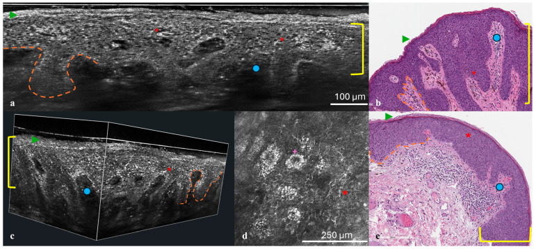
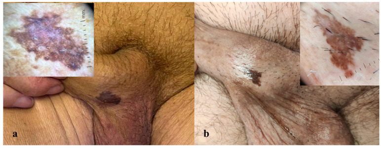
Penile intraepithelial neoplasia (PeIN) is a rare but clinically significant condition that can progress to invasive squamous carcinoma. Early diagnosis is crucial but often challenging due to its heterogeneous clinical and dermoscopic presentation, which can mimic other benign or malignant lesions. In this study, we report two cases of pigmented penile lesions evaluated using non-invasive imaging techniques: reflectance confocal microscopy (RCM) and line-field confocal optical coherence tomography (LC-OCT). Both methods revealed characteristic features such as hyperkeratosis, parakeratosis, acanthosis, nuclear pleomorphism of keratinocytes, and the presence of bright intraepithelial dendritic cells, correlating closely with histopathological findings of high-grade basaloid PeIN. Our findings highlight the valuable role of RCM and LC-OCT in improving the differential diagnosis of genital lesions, potentially reducing the need for invasive diagnostic procedures and ensuring early, appropriate management.

 求助内容:
求助内容: 应助结果提醒方式:


