Jin Ju Lee, Bo Ram Kwon, Min Young Lee, Ji Yeon Byun, Joo Young Roh, Hae Young Choi, You Won Choi
{"title":"No difference in inflammatory mediator expression between mast cell-rich and mast cell-poor rosacea lesions in Korean patients: a comparative study.","authors":"Jin Ju Lee, Bo Ram Kwon, Min Young Lee, Ji Yeon Byun, Joo Young Roh, Hae Young Choi, You Won Choi","doi":"10.12771/emj.2024.e78","DOIUrl":null,"url":null,"abstract":"<p><p><b>Objectives:</b> This study aimed to evaluate the correlation between mast cell (MC) density in rosacea-affected skin and the expression of key inflammatory mediators, including IL-6, TNF-α, and cathelicidin LL-37. By comparing lesions rich in MCs with those having fewer MCs, we sought to elucidate the role of MCs in the inflammatory mechanisms underlying rosacea pathogenesis. <b>Methods:</b> Specimens were collected from 20 patients diagnosed with rosacea who attended the outpatient clinic between 2008 and 2013. Each specimen underwent staining using hematoxylin/eosin, Giemsa, IL-6, LL-37, and TNF-α for both histopathological and immunohistochemical analyses. The number of stained cells was counted across 10 randomly selected dermal layers at a magnification of ×400 using light microscopy. The results were categorized based on the number of MCs counted: more than 10 MCs were classified as MC-rich, and 10 or fewer MCs as MC-poor. <b>Results:</b> Among the 20 patients (10 MC-rich and 10 MC-poor), the MC-rich group demonstrated significantly higher MC counts than the MC-poor group (P<0.001). However, there were no significant differences in the expression levels of IL-6, LL-37, or TNF-α between the two groups. Additionally, MC density did not show any significant associations with patient demographics, clinical characteristics, or systemic comorbidities. <b>Conclusion:</b> Increased MC density was not associated with differences in IL-6, TNF-α, or LL-37 expression in rosacea lesions. These findings suggest that MC infiltration may not directly influence the inflammatory mediator profile in rosacea. Further research is required to identify distinctive pathological features or markers that can elucidate the mechanisms of rosacea.</p>","PeriodicalId":41392,"journal":{"name":"Ewha Medical Journal","volume":"48 1","pages":"e78"},"PeriodicalIF":0.2000,"publicationDate":"2025-01-01","publicationTypes":"Journal Article","fieldsOfStudy":null,"isOpenAccess":false,"openAccessPdf":"https://www.ncbi.nlm.nih.gov/pmc/articles/PMC12277890/pdf/","citationCount":"0","resultStr":null,"platform":"Semanticscholar","paperid":null,"PeriodicalName":"Ewha Medical Journal","FirstCategoryId":"1085","ListUrlMain":"https://doi.org/10.12771/emj.2024.e78","RegionNum":0,"RegionCategory":null,"ArticlePicture":[],"TitleCN":null,"AbstractTextCN":null,"PMCID":null,"EPubDate":"2025/1/31 0:00:00","PubModel":"Epub","JCR":"Q3","JCRName":"MEDICINE, GENERAL & INTERNAL","Score":null,"Total":0}
引用次数: 0
Abstract
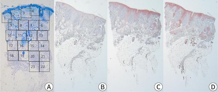
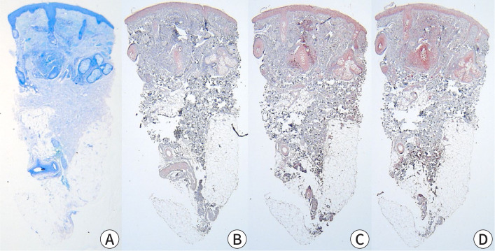
Objectives: This study aimed to evaluate the correlation between mast cell (MC) density in rosacea-affected skin and the expression of key inflammatory mediators, including IL-6, TNF-α, and cathelicidin LL-37. By comparing lesions rich in MCs with those having fewer MCs, we sought to elucidate the role of MCs in the inflammatory mechanisms underlying rosacea pathogenesis. Methods: Specimens were collected from 20 patients diagnosed with rosacea who attended the outpatient clinic between 2008 and 2013. Each specimen underwent staining using hematoxylin/eosin, Giemsa, IL-6, LL-37, and TNF-α for both histopathological and immunohistochemical analyses. The number of stained cells was counted across 10 randomly selected dermal layers at a magnification of ×400 using light microscopy. The results were categorized based on the number of MCs counted: more than 10 MCs were classified as MC-rich, and 10 or fewer MCs as MC-poor. Results: Among the 20 patients (10 MC-rich and 10 MC-poor), the MC-rich group demonstrated significantly higher MC counts than the MC-poor group (P<0.001). However, there were no significant differences in the expression levels of IL-6, LL-37, or TNF-α between the two groups. Additionally, MC density did not show any significant associations with patient demographics, clinical characteristics, or systemic comorbidities. Conclusion: Increased MC density was not associated with differences in IL-6, TNF-α, or LL-37 expression in rosacea lesions. These findings suggest that MC infiltration may not directly influence the inflammatory mediator profile in rosacea. Further research is required to identify distinctive pathological features or markers that can elucidate the mechanisms of rosacea.

 求助内容:
求助内容: 应助结果提醒方式:


