{"title":"Genotype-specific retinal and choroidal perfusion patterns in inherited retinal diseases: an SS-OCTA analysis.","authors":"Yu Rong, Junfeng Li, Jianquan He, Daowei Zhang, Jiawen Wu, Hongli Liu, Ting Li, Ping Xu, Qing Chang, Jihong Wu","doi":"10.1186/s40942-025-00706-0","DOIUrl":null,"url":null,"abstract":"<p><strong>Background: </strong>Retinitis pigmentosa (RP), an inherited retinal disease, is characterized by progressive vision loss driven by the gradual degeneration of retinal photoreceptors. This process manifests as impaired dark adaptation, night blindness, constriction of the visual field, and the deterioration of central vision. Although the progression can be monitored by electroretinography (ERG), visual field (VF) tests and optical coherence tomography (OCT) to some extent, it's hard to achieve high repeatability. Considering the correlation between patients' retinal blood volume and their visual function, OCT angiography (OCTA) can be a good choice for monitoring RP progression by objectively quantifying vascular changes.</p><p><strong>Methods: </strong>This study included 62 patients and 21 matched controls. Patients with RP were classified into five groups based on their genotype (CYP4V2, EYS, PRPH2, RPGR, and USH2A). Quantitative measurements and analyses were performed in nine fields of the fundus.</p><p><strong>Results: </strong>Defects were observed in each layer among all RP groups, showing different patterns of damage to the vasculature of the SCP, DCP, CC, and MLC. Foveal avascular zone (FAZ) sizes of the SCP and DCP in CYP4V2 and EYS groups, respectively, were larger than those in healthy individuals; PDs were associated with retinal function in each group. The CVI decreased to various degrees based on genotype and was associated with retinal function.</p><p><strong>Conclusion: </strong>Patients with RP had decreased PDs in the retina and choroid. PDs correlated with specific genotypes and retinal functions. SS-OCTA may be a non-invasive method for detecting the severity of RP.</p>","PeriodicalId":14289,"journal":{"name":"International Journal of Retina and Vitreous","volume":"11 1","pages":"82"},"PeriodicalIF":2.4000,"publicationDate":"2025-07-23","publicationTypes":"Journal Article","fieldsOfStudy":null,"isOpenAccess":false,"openAccessPdf":"https://www.ncbi.nlm.nih.gov/pmc/articles/PMC12288356/pdf/","citationCount":"0","resultStr":null,"platform":"Semanticscholar","paperid":null,"PeriodicalName":"International Journal of Retina and Vitreous","FirstCategoryId":"1085","ListUrlMain":"https://doi.org/10.1186/s40942-025-00706-0","RegionNum":0,"RegionCategory":null,"ArticlePicture":[],"TitleCN":null,"AbstractTextCN":null,"PMCID":null,"EPubDate":"","PubModel":"","JCR":"Q2","JCRName":"OPHTHALMOLOGY","Score":null,"Total":0}
引用次数: 0
Abstract
Background: Retinitis pigmentosa (RP), an inherited retinal disease, is characterized by progressive vision loss driven by the gradual degeneration of retinal photoreceptors. This process manifests as impaired dark adaptation, night blindness, constriction of the visual field, and the deterioration of central vision. Although the progression can be monitored by electroretinography (ERG), visual field (VF) tests and optical coherence tomography (OCT) to some extent, it's hard to achieve high repeatability. Considering the correlation between patients' retinal blood volume and their visual function, OCT angiography (OCTA) can be a good choice for monitoring RP progression by objectively quantifying vascular changes.
Methods: This study included 62 patients and 21 matched controls. Patients with RP were classified into five groups based on their genotype (CYP4V2, EYS, PRPH2, RPGR, and USH2A). Quantitative measurements and analyses were performed in nine fields of the fundus.
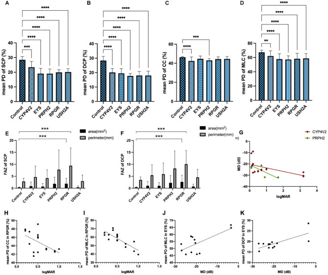
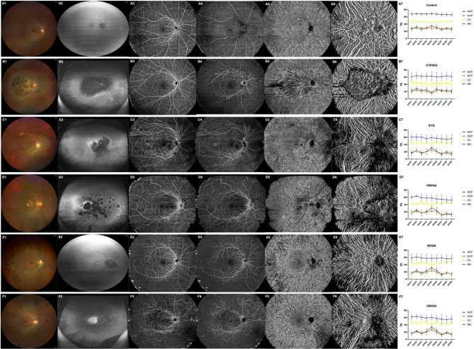
Results: Defects were observed in each layer among all RP groups, showing different patterns of damage to the vasculature of the SCP, DCP, CC, and MLC. Foveal avascular zone (FAZ) sizes of the SCP and DCP in CYP4V2 and EYS groups, respectively, were larger than those in healthy individuals; PDs were associated with retinal function in each group. The CVI decreased to various degrees based on genotype and was associated with retinal function.
Conclusion: Patients with RP had decreased PDs in the retina and choroid. PDs correlated with specific genotypes and retinal functions. SS-OCTA may be a non-invasive method for detecting the severity of RP.
期刊介绍:
International Journal of Retina and Vitreous focuses on the ophthalmic subspecialty of vitreoretinal disorders. The journal presents original articles on new approaches to diagnosis, outcomes of clinical trials, innovations in pharmacological therapy and surgical techniques, as well as basic science advances that impact clinical practice. Topical areas include, but are not limited to: -Imaging of the retina, choroid and vitreous -Innovations in optical coherence tomography (OCT) -Small-gauge vitrectomy, retinal detachment, chromovitrectomy -Electroretinography (ERG), microperimetry, other functional tests -Intraocular tumors -Retinal pharmacotherapy & drug delivery -Diabetic retinopathy & other vascular diseases -Age-related macular degeneration (AMD) & other macular entities

 求助内容:
求助内容: 应助结果提醒方式:


