Rizaldy Taslim Pinzon, Marlyna Afifudin, Ananda Digdoyo, Fx Kevin Christiansen Naibaho, Petra Gusti Parikesit, Yoel Sasamu Allendio
{"title":"Foster Kennedy Syndrome from Frontal Lobe Meningioma: A Rare Case Report.","authors":"Rizaldy Taslim Pinzon, Marlyna Afifudin, Ananda Digdoyo, Fx Kevin Christiansen Naibaho, Petra Gusti Parikesit, Yoel Sasamu Allendio","doi":"10.22336/rjo.2025.43","DOIUrl":null,"url":null,"abstract":"<p><strong>Background: </strong>Foster Kennedy syndrome is a neuro-ophthalmological disorder characterized by ipsilateral vision loss in one eye, followed by clinically significant papilledema in the opposite eye. The presence of mass lesions in the frontal lobe is primarily responsible for this syndrome. This case report further discusses symblepharon as an ocular manifestation of SJS.</p><p><strong>Method: </strong>A case report.</p><p><strong>Case report: </strong>We present a case of a 59-year-old female with a history of progressive headache, anosmia, mental status changes, and progressive poor vision. Ocular examination revealed disc pallor in her left eye with disc oedema in the contralateral eye. The patient was sent for computerized tomography (CT) and MRI, and the diagnosis of frontal lobe meningioma was confirmed. The surgical removal was performed, and the condition improved gradually.</p><p><strong>Discussion: </strong>We present a case of Foster Kennedy Syndrome, a rare neurological sign characterized by optic atrophy (vision loss) in one eye and papilledema (swelling of the optic nerve) in the other eye, often associated with an intracranial mass (meningioma).</p><p><strong>Conclusion: </strong>Presence of cranial fossa meningioma related to direct compression of a unilateral optic nerve, resulting in optic atrophy and might induce a rise in intracranial pressure, resulting in contralateral papilledema. This case presentation demonstrated that prompt and appropriate treatment was effective in gradually reducing the deterioration of symptoms associated with Foster Kennedy syndrome.</p>","PeriodicalId":94355,"journal":{"name":"Romanian journal of ophthalmology","volume":"69 2","pages":"271-274"},"PeriodicalIF":0.0000,"publicationDate":"2025-04-01","publicationTypes":"Journal Article","fieldsOfStudy":null,"isOpenAccess":false,"openAccessPdf":"https://www.ncbi.nlm.nih.gov/pmc/articles/PMC12277997/pdf/","citationCount":"0","resultStr":null,"platform":"Semanticscholar","paperid":null,"PeriodicalName":"Romanian journal of ophthalmology","FirstCategoryId":"1085","ListUrlMain":"https://doi.org/10.22336/rjo.2025.43","RegionNum":0,"RegionCategory":null,"ArticlePicture":[],"TitleCN":null,"AbstractTextCN":null,"PMCID":null,"EPubDate":"","PubModel":"","JCR":"","JCRName":"","Score":null,"Total":0}
引用次数: 0
Abstract
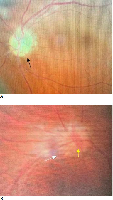
Background: Foster Kennedy syndrome is a neuro-ophthalmological disorder characterized by ipsilateral vision loss in one eye, followed by clinically significant papilledema in the opposite eye. The presence of mass lesions in the frontal lobe is primarily responsible for this syndrome. This case report further discusses symblepharon as an ocular manifestation of SJS.
Method: A case report.
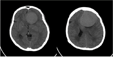
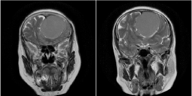
Case report: We present a case of a 59-year-old female with a history of progressive headache, anosmia, mental status changes, and progressive poor vision. Ocular examination revealed disc pallor in her left eye with disc oedema in the contralateral eye. The patient was sent for computerized tomography (CT) and MRI, and the diagnosis of frontal lobe meningioma was confirmed. The surgical removal was performed, and the condition improved gradually.
Discussion: We present a case of Foster Kennedy Syndrome, a rare neurological sign characterized by optic atrophy (vision loss) in one eye and papilledema (swelling of the optic nerve) in the other eye, often associated with an intracranial mass (meningioma).
Conclusion: Presence of cranial fossa meningioma related to direct compression of a unilateral optic nerve, resulting in optic atrophy and might induce a rise in intracranial pressure, resulting in contralateral papilledema. This case presentation demonstrated that prompt and appropriate treatment was effective in gradually reducing the deterioration of symptoms associated with Foster Kennedy syndrome.

 求助内容:
求助内容: 应助结果提醒方式:


