Jacky Huang, Banu Yagmurlu, Powell Molleti, Richard Lee, Abigail VanderPloeg, Humaira Noor, Rohan Bareja, Yiheng Li, Michael Iv, Haruka Itakura
{"title":"Brain tumor segmentation using deep learning: high performance with minimized MRI data.","authors":"Jacky Huang, Banu Yagmurlu, Powell Molleti, Richard Lee, Abigail VanderPloeg, Humaira Noor, Rohan Bareja, Yiheng Li, Michael Iv, Haruka Itakura","doi":"10.3389/fradi.2025.1616293","DOIUrl":null,"url":null,"abstract":"<p><strong>Purpose: </strong>Brain tumor segmentation with MRI is a challenging task, traditionally relying on manual delineation of regions-of-interest across multiple imaging sequences. However, this data-intensive approach is time-consuming. We aimed to optimize the process by using a deep learning (DL) based model while minimizing the number of MRI sequences required to segment gliomas.</p><p><strong>Methods: </strong>We trained a 3D U-Net DL model using the annotated 2018 MICCAI BraTS dataset (training dataset, <i>n</i> = 285), focusing on sub-segmenting enhancing tumor (ET) and tumor core (TC). We compared the performances of models trained on four different combinations of MRI sequences: T1C-only, FLAIR-only, T1C + FLAIR and T1 + T2 + T1C + FLAIR to evaluate whether a smaller MRI data subset could achieve comparable performance. We evaluated the performance on the four different sequence combinations using 5-fold cross-validation on the training dataset, then on our test dataset (<i>n</i> = 358) consisting of samples from a separately held-out 2018 BraTS validation set (<i>n</i> = 66) and 2021 BraTS datasets (<i>n</i> = 292). Dice scores on both cross-validation and test datasets were assessed to measure model performance.</p><p><strong>Results: </strong>Dice scores on cross-validation showed that T1C + FLAIR (ET: 0.814, TC: 0.856) matched or outperformed those of T1 + T2 + T1C + FLAIR (ET: 0.785, TC: 0.841), T1C-only (ET: 0.781, TC: 0.852) and FLAIR-only (ET: 0.008, TC: 0.619). Results on the test dataset also showed that T1C + FLAIR (ET: 0.867, TC: 0.926) matched or outperformed those of T1 + T2 + T1C + FLAIR (ET: 0.835, TC: 0.908), T1C-only (ET: 0.726, TC: 0.928), and FLAIR-only (ET: 0.056, TC: 0.543). T1C + FLAIR excelled in both ET and TC, exceeding the performance of the four-sequence dataset. T1C-only matched T1C + FLAIR in TC performance. Similarly<b>,</b> T1C and T1C + FLAIR also outperformed in ET delineation by sensitivity (0.829) and Hausdorff distance (5.964) on the test set. Across all configurations, specificity remained high (≥0.958). T1C performed well in TC delineation (sensitivity: 0.737), but the inclusion of all sequences led to improvement (0.754). Hausdorff distances clustered in a narrow range (17.622-33.812) for TC delineation across the configurations.</p><p><strong>Conclusions: </strong>DL-based brain tumor segmentation can achieve high accuracy using only two MRI sequences (T1C + FLAIR). Reduction of multiple sequence dependency may enhance DL generalizability and dissemination in both clinical and research contexts. Our findings may ultimately help mitigate human labor intensity of a complex task integral to medical imaging analysis.</p>","PeriodicalId":73101,"journal":{"name":"Frontiers in radiology","volume":"5 ","pages":"1616293"},"PeriodicalIF":2.3000,"publicationDate":"2025-07-08","publicationTypes":"Journal Article","fieldsOfStudy":null,"isOpenAccess":false,"openAccessPdf":"https://www.ncbi.nlm.nih.gov/pmc/articles/PMC12281592/pdf/","citationCount":"0","resultStr":null,"platform":"Semanticscholar","paperid":null,"PeriodicalName":"Frontiers in radiology","FirstCategoryId":"1085","ListUrlMain":"https://doi.org/10.3389/fradi.2025.1616293","RegionNum":0,"RegionCategory":null,"ArticlePicture":[],"TitleCN":null,"AbstractTextCN":null,"PMCID":null,"EPubDate":"2025/1/1 0:00:00","PubModel":"eCollection","JCR":"","JCRName":"","Score":null,"Total":0}
引用次数: 0
Abstract
Purpose: Brain tumor segmentation with MRI is a challenging task, traditionally relying on manual delineation of regions-of-interest across multiple imaging sequences. However, this data-intensive approach is time-consuming. We aimed to optimize the process by using a deep learning (DL) based model while minimizing the number of MRI sequences required to segment gliomas.
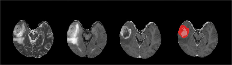
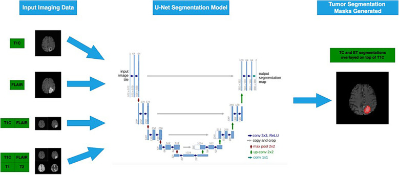
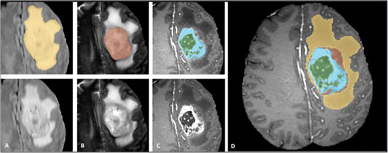
Methods: We trained a 3D U-Net DL model using the annotated 2018 MICCAI BraTS dataset (training dataset, n = 285), focusing on sub-segmenting enhancing tumor (ET) and tumor core (TC). We compared the performances of models trained on four different combinations of MRI sequences: T1C-only, FLAIR-only, T1C + FLAIR and T1 + T2 + T1C + FLAIR to evaluate whether a smaller MRI data subset could achieve comparable performance. We evaluated the performance on the four different sequence combinations using 5-fold cross-validation on the training dataset, then on our test dataset (n = 358) consisting of samples from a separately held-out 2018 BraTS validation set (n = 66) and 2021 BraTS datasets (n = 292). Dice scores on both cross-validation and test datasets were assessed to measure model performance.
Results: Dice scores on cross-validation showed that T1C + FLAIR (ET: 0.814, TC: 0.856) matched or outperformed those of T1 + T2 + T1C + FLAIR (ET: 0.785, TC: 0.841), T1C-only (ET: 0.781, TC: 0.852) and FLAIR-only (ET: 0.008, TC: 0.619). Results on the test dataset also showed that T1C + FLAIR (ET: 0.867, TC: 0.926) matched or outperformed those of T1 + T2 + T1C + FLAIR (ET: 0.835, TC: 0.908), T1C-only (ET: 0.726, TC: 0.928), and FLAIR-only (ET: 0.056, TC: 0.543). T1C + FLAIR excelled in both ET and TC, exceeding the performance of the four-sequence dataset. T1C-only matched T1C + FLAIR in TC performance. Similarly, T1C and T1C + FLAIR also outperformed in ET delineation by sensitivity (0.829) and Hausdorff distance (5.964) on the test set. Across all configurations, specificity remained high (≥0.958). T1C performed well in TC delineation (sensitivity: 0.737), but the inclusion of all sequences led to improvement (0.754). Hausdorff distances clustered in a narrow range (17.622-33.812) for TC delineation across the configurations.
Conclusions: DL-based brain tumor segmentation can achieve high accuracy using only two MRI sequences (T1C + FLAIR). Reduction of multiple sequence dependency may enhance DL generalizability and dissemination in both clinical and research contexts. Our findings may ultimately help mitigate human labor intensity of a complex task integral to medical imaging analysis.

 求助内容:
求助内容: 应助结果提醒方式:


