Veer Sangha, Lovedeep Singh Dhingra, Arya Aminorroaya, Philip M. Croon, Nikhil V. Sikand, Sounok Sen, Matthew W. Martinez, Martin S. Maron, Harlan M. Krumholz, Folkert W. Asselbergs, Evangelos K. Oikonomou, Rohan Khera
{"title":"Identification of hypertrophic cardiomyopathy on electrocardiographic images with deep learning","authors":"Veer Sangha, Lovedeep Singh Dhingra, Arya Aminorroaya, Philip M. Croon, Nikhil V. Sikand, Sounok Sen, Matthew W. Martinez, Martin S. Maron, Harlan M. Krumholz, Folkert W. Asselbergs, Evangelos K. Oikonomou, Rohan Khera","doi":"10.1038/s44161-025-00685-3","DOIUrl":null,"url":null,"abstract":"Hypertrophic cardiomyopathy (HCM) is frequently underdiagnosed. Although deep learning (DL) models using raw electrocardiographic (ECG) voltage data can enhance detection, their use at the point of care is limited. Here we report the development and validation of a DL model that detects HCM from images of 12-lead ECGs across layouts. The model was developed using 124,553 ECGs from 66,987 individuals at the Yale New Haven Hospital (YNHH), with HCM features determined by concurrent imaging (cardiac magnetic resonance (CMR) or echocardiography). External validation included ECG images from MIMIC-IV, the Amsterdam University Medical Center (AUMC) and the UK Biobank (UKB), where HCM was defined by CMR (YNHH, MIMIC-IV and AUMC) and diagnosis codes (UKB). The model demonstrated robust performance across image formats and sites (areas under the receiver operating characteristic curve (AUROCs): 0.95 internal testing; 0.94 MIMIC-IV; 0.92 AUMC; 0.91 UKB). Discriminative features localized to anterior/lateral leads (V4 and V5) regardless of layout. This approach enables scalable, image-based screening for HCM across clinical settings. Sangha, Dhingra et al. develop and validate a deep learning model to diagnose hypertrophic cardiomyopathy from electrocardiographic images, demonstrating its effectiveness across multiple 12-lead layouts.","PeriodicalId":74245,"journal":{"name":"Nature cardiovascular research","volume":"4 8","pages":"991-1000"},"PeriodicalIF":10.8000,"publicationDate":"2025-07-22","publicationTypes":"Journal Article","fieldsOfStudy":null,"isOpenAccess":false,"openAccessPdf":"","citationCount":"0","resultStr":null,"platform":"Semanticscholar","paperid":null,"PeriodicalName":"Nature cardiovascular research","FirstCategoryId":"1085","ListUrlMain":"https://www.nature.com/articles/s44161-025-00685-3","RegionNum":0,"RegionCategory":null,"ArticlePicture":[],"TitleCN":null,"AbstractTextCN":null,"PMCID":null,"EPubDate":"","PubModel":"","JCR":"Q1","JCRName":"CARDIAC & CARDIOVASCULAR SYSTEMS","Score":null,"Total":0}
引用次数: 0
Abstract
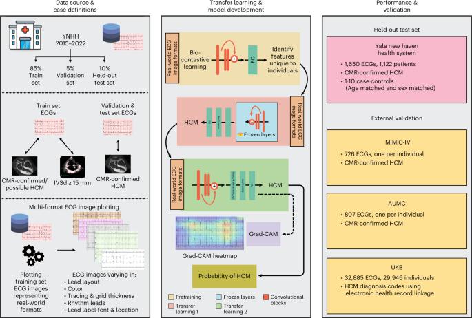
Hypertrophic cardiomyopathy (HCM) is frequently underdiagnosed. Although deep learning (DL) models using raw electrocardiographic (ECG) voltage data can enhance detection, their use at the point of care is limited. Here we report the development and validation of a DL model that detects HCM from images of 12-lead ECGs across layouts. The model was developed using 124,553 ECGs from 66,987 individuals at the Yale New Haven Hospital (YNHH), with HCM features determined by concurrent imaging (cardiac magnetic resonance (CMR) or echocardiography). External validation included ECG images from MIMIC-IV, the Amsterdam University Medical Center (AUMC) and the UK Biobank (UKB), where HCM was defined by CMR (YNHH, MIMIC-IV and AUMC) and diagnosis codes (UKB). The model demonstrated robust performance across image formats and sites (areas under the receiver operating characteristic curve (AUROCs): 0.95 internal testing; 0.94 MIMIC-IV; 0.92 AUMC; 0.91 UKB). Discriminative features localized to anterior/lateral leads (V4 and V5) regardless of layout. This approach enables scalable, image-based screening for HCM across clinical settings. Sangha, Dhingra et al. develop and validate a deep learning model to diagnose hypertrophic cardiomyopathy from electrocardiographic images, demonstrating its effectiveness across multiple 12-lead layouts.

 求助内容:
求助内容: 应助结果提醒方式:


