{"title":"Treatment of lumbar tuberculosis with minimally invasive anterior lesion clearance combined with posterior fixation.","authors":"Fei-Fei Pu, Xiang-Lin Peng, Fang-Zheng Zhou, Xiao-Long Zhao, Ling Yang, Jun-Qing Cao, Liu Wei, Jing Feng, Ping Xia","doi":"10.5312/wjo.v16.i7.106041","DOIUrl":null,"url":null,"abstract":"<p><strong>Background: </strong>Spinal tuberculosis, a destructive extrapulmonary form, often causes severe deformity and neurological deficits. Surgical intervention aims to debride lesions, reconstruct stability, and correct deformities. This study evaluates a combined posterior fixation and minimally invasive anterior approach for lumbar tuberculosis.</p><p><strong>Aim: </strong>To evaluate the clinical outcomes and radiological parameters of posterior internal fixation combined with minimally invasive anterior lesion clearance and bone graft fusion for the treatment of lumbar tuberculosis.</p><p><strong>Methods: </strong>Clinical data from 24 patients with lumbar tuberculosis who underwent posterior pedicle screw fixation combined with minimally invasive anterior lesion clearance were analyzed. The Cobb angle, visual analog scale (VAS) score, and Frankel classification were statistically assessed preoperatively and postoperatively. Complications and bone graft fusion were also recorded.</p><p><strong>Results: </strong>Wounds healed in the first stage in 22 patients; one patient developed a posterior incisional sinus tract, and one experienced postoperative tuberculosis recurrence. At the final follow-up, according to the Frankel classification, there were 1, 2, and 21 cases classified as grade C, grade D, and grade E, respectively. By the last follow-up, the Cobb angle, VAS score, and erythrocyte sedimentation rate had all decreased. Both X-ray and computed tomography images confirmed bone healing. The fusion time ranged from 3 to 9 months, with an average of 5.2 months.</p><p><strong>Conclusion: </strong>Posterior pedicle screw fixation combined with minimally invasive anterior lesion clearance is an effective and safe treatment for lumbar tuberculosis.</p>","PeriodicalId":47843,"journal":{"name":"World Journal of Orthopedics","volume":"16 7","pages":"106041"},"PeriodicalIF":2.3000,"publicationDate":"2025-07-18","publicationTypes":"Journal Article","fieldsOfStudy":null,"isOpenAccess":false,"openAccessPdf":"https://www.ncbi.nlm.nih.gov/pmc/articles/PMC12278289/pdf/","citationCount":"0","resultStr":null,"platform":"Semanticscholar","paperid":null,"PeriodicalName":"World Journal of Orthopedics","FirstCategoryId":"1085","ListUrlMain":"https://doi.org/10.5312/wjo.v16.i7.106041","RegionNum":0,"RegionCategory":null,"ArticlePicture":[],"TitleCN":null,"AbstractTextCN":null,"PMCID":null,"EPubDate":"","PubModel":"","JCR":"Q2","JCRName":"ORTHOPEDICS","Score":null,"Total":0}
引用次数: 0
Abstract
Background: Spinal tuberculosis, a destructive extrapulmonary form, often causes severe deformity and neurological deficits. Surgical intervention aims to debride lesions, reconstruct stability, and correct deformities. This study evaluates a combined posterior fixation and minimally invasive anterior approach for lumbar tuberculosis.
Aim: To evaluate the clinical outcomes and radiological parameters of posterior internal fixation combined with minimally invasive anterior lesion clearance and bone graft fusion for the treatment of lumbar tuberculosis.
Methods: Clinical data from 24 patients with lumbar tuberculosis who underwent posterior pedicle screw fixation combined with minimally invasive anterior lesion clearance were analyzed. The Cobb angle, visual analog scale (VAS) score, and Frankel classification were statistically assessed preoperatively and postoperatively. Complications and bone graft fusion were also recorded.
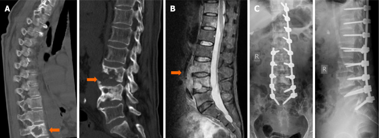
Results: Wounds healed in the first stage in 22 patients; one patient developed a posterior incisional sinus tract, and one experienced postoperative tuberculosis recurrence. At the final follow-up, according to the Frankel classification, there were 1, 2, and 21 cases classified as grade C, grade D, and grade E, respectively. By the last follow-up, the Cobb angle, VAS score, and erythrocyte sedimentation rate had all decreased. Both X-ray and computed tomography images confirmed bone healing. The fusion time ranged from 3 to 9 months, with an average of 5.2 months.
Conclusion: Posterior pedicle screw fixation combined with minimally invasive anterior lesion clearance is an effective and safe treatment for lumbar tuberculosis.

 求助内容:
求助内容: 应助结果提醒方式:


