{"title":"Rupture of Life-Threatening Hepatic Artery Pseudoaneurysm After Endoscopic Ultrasonography-guided Hepaticogastrostomy: Successful Management With Emergency Transcatheter Arterial Embolization","authors":"Hiroshi Yukimoto, Akino Okamoto, Kohsaku Ohnishi, Keitaro Masuko, Junping Wang, Kazuya Ogawa, Ken Ueda, Motohiro Hirao, Yasuhiro Nakaya, Atsushi Hosui","doi":"10.1002/deo2.70176","DOIUrl":null,"url":null,"abstract":"<p>A 70-year-old male with lung cancer and interstitial pneumonia was diagnosed with ampullary carcinoma, causing obstructive jaundice. After the failure of endoscopic retrograde cholangiopancreatography, endoscopic ultrasound-guided hepaticogastrostomy (EUS-HGS) was performed with a 7-Fr plastic stent (PS) into the B2 bile duct. Three months later, mild bleeding was observed during stent exchange, but was stopped by stent replacement. The patient developed recurrent cholangitis, and 1 month later, when the PS was removed to add supplementary drainage, massive bleeding occurred from the endosonographically created route into the stomach. Contrast-enhanced computed tomography (CECT) revealed a pseudoaneurysm in the A2 branch of the hepatic artery. Emergency angiography confirmed active extravasation, and successful transcatheter arterial embolization with <i>N</i>-butyl-2-cyanoacrylate was performed. The patient recovered without rebleeding but died two weeks later from worsening interstitial pneumonia. A review of publications identified only three previous cases of pseudoaneurysm after EUS-HGS, all of which involved self-expandable metal stents. This case demonstrates that pseudoaneurysms can cause both gastrointestinal bleeding and recurrent cholangitis. Careful evaluation of CECT images is needed before stent manipulation in patients with biliary symptoms after EUS-HGS.</p>","PeriodicalId":93973,"journal":{"name":"DEN open","volume":"6 1","pages":""},"PeriodicalIF":1.5000,"publicationDate":"2025-07-24","publicationTypes":"Journal Article","fieldsOfStudy":null,"isOpenAccess":false,"openAccessPdf":"https://onlinelibrary.wiley.com/doi/epdf/10.1002/deo2.70176","citationCount":"0","resultStr":null,"platform":"Semanticscholar","paperid":null,"PeriodicalName":"DEN open","FirstCategoryId":"1085","ListUrlMain":"https://onlinelibrary.wiley.com/doi/10.1002/deo2.70176","RegionNum":0,"RegionCategory":null,"ArticlePicture":[],"TitleCN":null,"AbstractTextCN":null,"PMCID":null,"EPubDate":"","PubModel":"","JCR":"Q4","JCRName":"GASTROENTEROLOGY & HEPATOLOGY","Score":null,"Total":0}
引用次数: 0
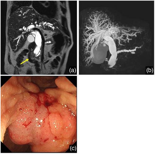
Abstract
A 70-year-old male with lung cancer and interstitial pneumonia was diagnosed with ampullary carcinoma, causing obstructive jaundice. After the failure of endoscopic retrograde cholangiopancreatography, endoscopic ultrasound-guided hepaticogastrostomy (EUS-HGS) was performed with a 7-Fr plastic stent (PS) into the B2 bile duct. Three months later, mild bleeding was observed during stent exchange, but was stopped by stent replacement. The patient developed recurrent cholangitis, and 1 month later, when the PS was removed to add supplementary drainage, massive bleeding occurred from the endosonographically created route into the stomach. Contrast-enhanced computed tomography (CECT) revealed a pseudoaneurysm in the A2 branch of the hepatic artery. Emergency angiography confirmed active extravasation, and successful transcatheter arterial embolization with N-butyl-2-cyanoacrylate was performed. The patient recovered without rebleeding but died two weeks later from worsening interstitial pneumonia. A review of publications identified only three previous cases of pseudoaneurysm after EUS-HGS, all of which involved self-expandable metal stents. This case demonstrates that pseudoaneurysms can cause both gastrointestinal bleeding and recurrent cholangitis. Careful evaluation of CECT images is needed before stent manipulation in patients with biliary symptoms after EUS-HGS.

 求助内容:
求助内容: 应助结果提醒方式:


