Developmental Defect of Enamel in Permanent Teeth Associated With Chronic Endodontic Abscess in Deciduous Teeth: A Retrospective Study
Abstract
Objectives
Destructive carious lesions on deciduous teeth often result in dental abscesses. Sometimes, the exudative process may extend to the dental follicle of the permanent tooth, leading to various types of consequences. This study primarily seeks to determine the prevalence of developmental defects of enamel (DDE) in premolars whose predecessors developed endodontic abscesses. Furthermore, it investigates how the prevalence of DDE is influenced by the type of treatment the affected deciduous molar received. Lastly, the study compares the prevalence of DDE between maxillary and mandibular premolars.
Material and Methods
Demographics, medical and dental history, and records of DDE were extracted from the medical records of 1164 pediatric patients. DDE of 107 premolars from patients who had experienced abscesses in their deciduous molars were compared to DDE of 107 premolars from patients who naturally shed healthy deciduous molars. DDE were also compared between different treatment modalities and anatomical regions. Fisher's exact tests were used to compare groups, while demographic data were analyzed by descriptive statistics and reported as mean ± standard deviation or as median and interquartile range for the continuous variables.
Results
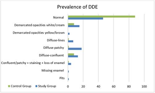
Compared to premolars whose predecessors did not exhibit signs of pathology, those that developed endodontic abscesses reported a higher prevalence of DDE (57% vs. 17.8%; OR 6.14; p < 0.0001). Endodontic treatment on deciduous molars was associated with higher DDE prevalence compared to surgical treatment (70.2% vs. 46.7%; OR 2.69; p = 0.016). Maxillary premolars showed a higher prevalence of DDE compared to mandibular premolars (75.4% vs. 24.6%; OR 5.23; p = 0.00008).
Conclusions
Chronic endodontic abscess on deciduous molars significantly increases the risk of DDE in the corresponding premolars. ET on deciduous molars is associated with higher incidence of DDE compared to extraction. Maxillary premolars are more likely to develop DDE than mandibular premolars.

 求助内容:
求助内容: 应助结果提醒方式:


