AI assisted diagnosis using DEANet to improve correct diagnosis of iliac wing fracture and ischial spine fracture
IF 1.4
3区 医学
Q4 ENGINEERING, BIOMEDICAL
引用次数: 0
Abstract
Background
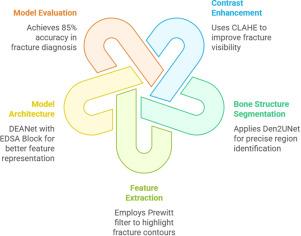
Hip fractures—particularly those involving the iliac wing and ischial spine—are complex injuries that often require CT or MRI scans for accurate diagnosis. However, these imaging modalities are costly, time-consuming, and involve exposure to radiation or contrast-related risks. This study aims to develop an AI-based classification model, the Dynamic Efficient Attention Network, capable of automatically distinguishing between iliac wing and ischial spine fractures using pelvic X-ray images. The objective is to facilitate early diagnosis and reduce reliance on advanced imaging modalities.
Methods
The proposed method employs a dual-branch architecture that integrates EfficientNet-B0 with an Enhanced Depth-wise Separable Attention Block to enhance edge-region feature representation. The model was trained using pelvic X-ray images collected from China Medical University Hospital and evaluated based on accuracy, precision, recall, F1 score, and Intersection over Union.
Findings
The model achieved an accuracy of 85 % on the test dataset and demonstrated robust performance across all evaluation metrics. These findings suggest that the proposed method has the potential to function as a reliable AI-assisted diagnostic tool for the early and accurate classification of hip fractures, thereby supporting clinical decision-making and improving treatment planning.
Interpretation
Compared to existing approaches that rely on CT or MRI imaging, the proposed method demonstrates that advanced processing of X-ray images can yield clinically meaningful classification results. This underscores the potential of the proposed method as a cost-effective, efficient, and accessible diagnostic tool, especially in settings where access to advanced imaging modalities is limited.
应用DEANet进行人工智能辅助诊断,提高髂翼骨折和坐骨棘骨折的正确诊断
背景:髋部骨折——尤其是涉及髂翼和坐骨棘的骨折——是一种复杂的损伤,通常需要CT或MRI扫描才能准确诊断。然而,这些成像方式是昂贵的,耗时的,并涉及暴露于辐射或对比度相关的风险。本研究旨在开发一种基于人工智能的分类模型——动态高效注意网络(Dynamic Efficient Attention Network),该模型能够利用骨盆x线图像自动区分髂翼骨折和坐骨棘骨折。目的是促进早期诊断,减少对先进成像方式的依赖。方法采用双分支结构,结合高效网络- b0和增强的深度可分离注意块,增强边缘区域特征表示。使用从中国医科大学附属医院收集的骨盆x线图像对该模型进行训练,并根据准确性、精密度、召回率、F1评分和交叉优于联合进行评估。该模型在测试数据集中实现了85%的准确性,并在所有评估指标中表现出稳健的性能。这些发现表明,所提出的方法有可能作为一种可靠的人工智能辅助诊断工具,用于髋部骨折的早期准确分类,从而支持临床决策和改进治疗计划。与现有的依赖于CT或MRI成像的方法相比,本文提出的方法表明,对x射线图像进行高级处理可以产生具有临床意义的分类结果。这强调了所提出的方法作为一种具有成本效益、效率高和可获得的诊断工具的潜力,特别是在获得先进成像模式的环境中。
本文章由计算机程序翻译,如有差异,请以英文原文为准。
求助全文
约1分钟内获得全文
求助全文
来源期刊

Clinical Biomechanics
医学-工程:生物医学
CiteScore
3.30
自引率
5.60%
发文量
189
审稿时长
12.3 weeks
期刊介绍:
Clinical Biomechanics is an international multidisciplinary journal of biomechanics with a focus on medical and clinical applications of new knowledge in the field.
The science of biomechanics helps explain the causes of cell, tissue, organ and body system disorders, and supports clinicians in the diagnosis, prognosis and evaluation of treatment methods and technologies. Clinical Biomechanics aims to strengthen the links between laboratory and clinic by publishing cutting-edge biomechanics research which helps to explain the causes of injury and disease, and which provides evidence contributing to improved clinical management.
A rigorous peer review system is employed and every attempt is made to process and publish top-quality papers promptly.
Clinical Biomechanics explores all facets of body system, organ, tissue and cell biomechanics, with an emphasis on medical and clinical applications of the basic science aspects. The role of basic science is therefore recognized in a medical or clinical context. The readership of the journal closely reflects its multi-disciplinary contents, being a balance of scientists, engineers and clinicians.
The contents are in the form of research papers, brief reports, review papers and correspondence, whilst special interest issues and supplements are published from time to time.
Disciplines covered include biomechanics and mechanobiology at all scales, bioengineering and use of tissue engineering and biomaterials for clinical applications, biophysics, as well as biomechanical aspects of medical robotics, ergonomics, physical and occupational therapeutics and rehabilitation.
 求助内容:
求助内容: 应助结果提醒方式:


