Hans-Jonas Meyer, Jakob Leonhardi, Anne-Kathrin Höhn, Noura Kabbani, Silke Zimmermann, Jan Borggrefe, Alexey Surov
{"title":"Associations between MRI radiomics analysis and tumor-micro milieu in uterine cervical cancer.","authors":"Hans-Jonas Meyer, Jakob Leonhardi, Anne-Kathrin Höhn, Noura Kabbani, Silke Zimmermann, Jan Borggrefe, Alexey Surov","doi":"10.1007/s00432-025-06253-3","DOIUrl":null,"url":null,"abstract":"<p><strong>Purpose: </strong>The complex interactions of the tumor micromilieu could be reflected by magnetic resonance imaging (MRI) when analyzed with the radiomics approach. For several tumor entities, it has been shown that radiomics derived from MRI can reflect important characteristics of the tumors. The present study investigated the association radiomics derived from MRI images and histopathological features in uterine cervical cancer.</p><p><strong>Methods: </strong>The MRI before any treatment was used to extract the radiomics features of T1- and T2-weighted images. The biopsy specimens were stained for Ki 67, e-cadherin, vimentin, programmed-death ligand 1, and tumor-infiltrating lymphocytes (TIL, all CD45 positive cells). Tumor-stroma ratio (TSR) was calculated on routine H&E specimen. Spearman's correlation analysis and discrimination analyses were performed as statistical analyses.</p><p><strong>Results: </strong>The patient sample was comprised of 89 female patients with a mean age of 49.3 years ± 14.6 (range 27-77 years) with squamous cell cervical carcinoma. \"Kurtosis\" derived from T1-weighted images after contrast media application correlated with the Ki-67 index (r = 0.28, p = 0.02). \"WavEnHL_s-4\" derived from T2-weighted images and \"S(1.0)Contrast\" derived from T1-weighted images after contrast media application showed correlations with TSR (r = - 0.24, p = 0.04, each). Several associations were identified between the radiomics features with immune scores defined by programmed-death ligand 1, the highest correlation showed Teta1 derived from T2-weighted images with the combined positive score (r = - 0.38, p < 0.01). There were several associations with vimentin expression, the highest showed \"Variance\" derived from T1-weighted images after contrast media application (r = 0.46, p < 0.01).</p><p><strong>Conclusions: </strong>Radiomics features derived from MRI can reflect tumor characteristics of UCC. Especially immune-related features were reflected by the MRI texture features. Proliferation potential, composition of the extracellular matrix and tumor-stroma ratio were also significantly associated with radiomics features. These presented results need to be evaluated in an independent cohort to test their stability.</p>","PeriodicalId":15118,"journal":{"name":"Journal of Cancer Research and Clinical Oncology","volume":"151 7","pages":"219"},"PeriodicalIF":2.8000,"publicationDate":"2025-07-21","publicationTypes":"Journal Article","fieldsOfStudy":null,"isOpenAccess":false,"openAccessPdf":"https://www.ncbi.nlm.nih.gov/pmc/articles/PMC12279667/pdf/","citationCount":"0","resultStr":null,"platform":"Semanticscholar","paperid":null,"PeriodicalName":"Journal of Cancer Research and Clinical Oncology","FirstCategoryId":"3","ListUrlMain":"https://doi.org/10.1007/s00432-025-06253-3","RegionNum":3,"RegionCategory":"医学","ArticlePicture":[],"TitleCN":null,"AbstractTextCN":null,"PMCID":null,"EPubDate":"","PubModel":"","JCR":"Q3","JCRName":"ONCOLOGY","Score":null,"Total":0}
引用次数: 0
Abstract
Purpose: The complex interactions of the tumor micromilieu could be reflected by magnetic resonance imaging (MRI) when analyzed with the radiomics approach. For several tumor entities, it has been shown that radiomics derived from MRI can reflect important characteristics of the tumors. The present study investigated the association radiomics derived from MRI images and histopathological features in uterine cervical cancer.
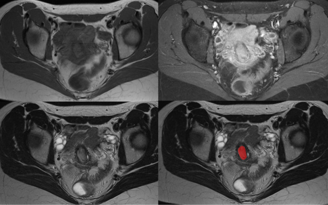
Methods: The MRI before any treatment was used to extract the radiomics features of T1- and T2-weighted images. The biopsy specimens were stained for Ki 67, e-cadherin, vimentin, programmed-death ligand 1, and tumor-infiltrating lymphocytes (TIL, all CD45 positive cells). Tumor-stroma ratio (TSR) was calculated on routine H&E specimen. Spearman's correlation analysis and discrimination analyses were performed as statistical analyses.
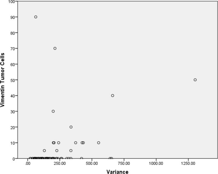
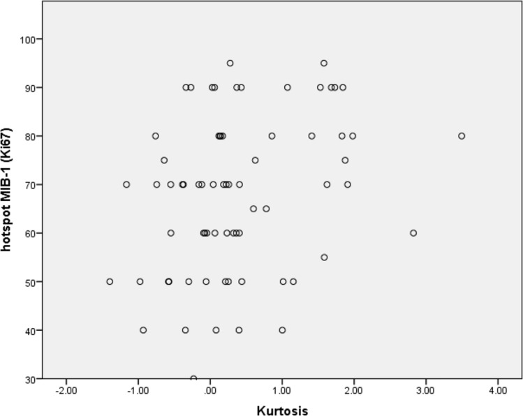
Results: The patient sample was comprised of 89 female patients with a mean age of 49.3 years ± 14.6 (range 27-77 years) with squamous cell cervical carcinoma. "Kurtosis" derived from T1-weighted images after contrast media application correlated with the Ki-67 index (r = 0.28, p = 0.02). "WavEnHL_s-4" derived from T2-weighted images and "S(1.0)Contrast" derived from T1-weighted images after contrast media application showed correlations with TSR (r = - 0.24, p = 0.04, each). Several associations were identified between the radiomics features with immune scores defined by programmed-death ligand 1, the highest correlation showed Teta1 derived from T2-weighted images with the combined positive score (r = - 0.38, p < 0.01). There were several associations with vimentin expression, the highest showed "Variance" derived from T1-weighted images after contrast media application (r = 0.46, p < 0.01).
Conclusions: Radiomics features derived from MRI can reflect tumor characteristics of UCC. Especially immune-related features were reflected by the MRI texture features. Proliferation potential, composition of the extracellular matrix and tumor-stroma ratio were also significantly associated with radiomics features. These presented results need to be evaluated in an independent cohort to test their stability.
期刊介绍:
The "Journal of Cancer Research and Clinical Oncology" publishes significant and up-to-date articles within the fields of experimental and clinical oncology. The journal, which is chiefly devoted to Original papers, also includes Reviews as well as Editorials and Guest editorials on current, controversial topics. The section Letters to the editors provides a forum for a rapid exchange of comments and information concerning previously published papers and topics of current interest. Meeting reports provide current information on the latest results presented at important congresses.
The following fields are covered: carcinogenesis - etiology, mechanisms; molecular biology; recent developments in tumor therapy; general diagnosis; laboratory diagnosis; diagnostic and experimental pathology; oncologic surgery; and epidemiology.
 求助内容:
求助内容: 应助结果提醒方式:


