{"title":"Internal and marginal adaptation of 3D printed interim fixed partial dentures with different layer thicknesses.","authors":"Fateme Ghorbanpour Arani, Mahya Hasanzade, Negin Aminianpour, Azam Sadat Mostafavi","doi":"10.4047/jap.2025.17.3.137","DOIUrl":null,"url":null,"abstract":"<p><strong>Purpose: </strong>The aim of this study was to compare the marginal and internal adaptation of polymethyl methacrylate interim fixed partial denture (FPD) restorations fabricated by three-dimensional (3D) printing with different layer thicknesses.</p><p><strong>Materials and methods: </strong>A standard typodont was scanned by a laboratory scanner. The maxillary left first premolar and first molar teeth received an all-ceramic full-coverage crown preparation, and the second premolar was removed to create a pontic space. The prepared teeth were then scanned, and metal dies were fabricated by a milling machine. Restorations were designed and fabricated in 3 groups (n = 12 in inch) with 25, 50 and 100 µm layer thicknesses. The marginal and internal gaps were measured by the replica technique. Data were analyzed by one-way ANOVA (α = 0.05).</p><p><strong>Results: </strong>The mean gap size (MGS) in the cervical area of premolar restorations was significantly higher in 100 µm than in 50 and 25 µm thicknesses (<i>P</i> < .05). The MGS in the marginal area of premolar restorations was significantly higher in 100 µm thickness than in 50 µm thickness (<i>P</i> < .05), with no statistically significant difference with 25 µm thickness (<i>P</i> > .05). The MGS at the occlusal and cervical areas of the molar restorations was significantly higher in 100 µm thickness than in 50 µm thickness (<i>P</i> < .05), with no statistically significant difference with 25 µm thickness (<i>P</i> > .05).</p><p><strong>Conclusion: </strong>Different thicknesses of additive layers in 3D printing affected the marginal and internal gaps. The smallest gap size was recorded in 50 µm layer thickness.</p>","PeriodicalId":51291,"journal":{"name":"Journal of Advanced Prosthodontics","volume":"17 3","pages":"137-145"},"PeriodicalIF":2.5000,"publicationDate":"2025-06-01","publicationTypes":"Journal Article","fieldsOfStudy":null,"isOpenAccess":false,"openAccessPdf":"https://www.ncbi.nlm.nih.gov/pmc/articles/PMC12270714/pdf/","citationCount":"0","resultStr":null,"platform":"Semanticscholar","paperid":null,"PeriodicalName":"Journal of Advanced Prosthodontics","FirstCategoryId":"3","ListUrlMain":"https://doi.org/10.4047/jap.2025.17.3.137","RegionNum":3,"RegionCategory":"医学","ArticlePicture":[],"TitleCN":null,"AbstractTextCN":null,"PMCID":null,"EPubDate":"2025/6/25 0:00:00","PubModel":"Epub","JCR":"Q1","JCRName":"DENTISTRY, ORAL SURGERY & MEDICINE","Score":null,"Total":0}
引用次数: 0
Abstract
Purpose: The aim of this study was to compare the marginal and internal adaptation of polymethyl methacrylate interim fixed partial denture (FPD) restorations fabricated by three-dimensional (3D) printing with different layer thicknesses.
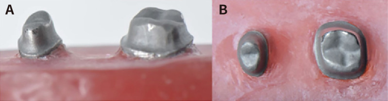
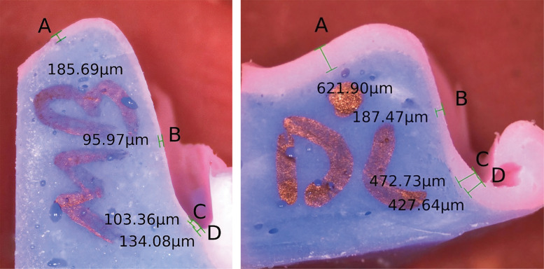
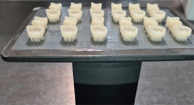
Materials and methods: A standard typodont was scanned by a laboratory scanner. The maxillary left first premolar and first molar teeth received an all-ceramic full-coverage crown preparation, and the second premolar was removed to create a pontic space. The prepared teeth were then scanned, and metal dies were fabricated by a milling machine. Restorations were designed and fabricated in 3 groups (n = 12 in inch) with 25, 50 and 100 µm layer thicknesses. The marginal and internal gaps were measured by the replica technique. Data were analyzed by one-way ANOVA (α = 0.05).
Results: The mean gap size (MGS) in the cervical area of premolar restorations was significantly higher in 100 µm than in 50 and 25 µm thicknesses (P < .05). The MGS in the marginal area of premolar restorations was significantly higher in 100 µm thickness than in 50 µm thickness (P < .05), with no statistically significant difference with 25 µm thickness (P > .05). The MGS at the occlusal and cervical areas of the molar restorations was significantly higher in 100 µm thickness than in 50 µm thickness (P < .05), with no statistically significant difference with 25 µm thickness (P > .05).
Conclusion: Different thicknesses of additive layers in 3D printing affected the marginal and internal gaps. The smallest gap size was recorded in 50 µm layer thickness.
期刊介绍:
This journal aims to convey scientific and clinical progress in the field of prosthodontics and its related areas to many dental communities concerned with esthetic and functional restorations, occlusion, implants, prostheses, and biomaterials related to prosthodontics.
This journal publishes
• Original research data of high scientific merit in the field of diagnosis, function, esthetics and stomatognathic physiology related to prosthodontic rehabilitation, physiology and mechanics of occlusion, mechanical and biologic aspects of prosthodontic materials including dental implants.
• Review articles by experts on controversies and new developments in prosthodontics.
• Case reports if they provide or document new fundamental knowledge.
 求助内容:
求助内容: 应助结果提醒方式:


