{"title":"CT-Based Evaluation of Bone Mineral Density Distribution of Proximal Femur in Patients With Femoral Trochanteric Fracture.","authors":"Daisuke Enomoto, Hyonmin Choe, Masahiro Matsumoto, Koki Abe, Kazuyoshi Yamamoto, Kousuke Matsuo, Hiroyuki Makita, Naomi Kobayashi, Yutaka Inaba","doi":"10.1177/21514593251361803","DOIUrl":null,"url":null,"abstract":"<p><strong>Background: </strong>Surgical treatment of proximal femoral fractures typically involves fixation with intramedullary nailing or sliding hip screws, where screws inserted into the femoral head stabilize the fracture site. However, few studies have quantitatively assessed the distribution of bone density and quality within the femoral head. We investigated the distribution of bone mineral density (BMD) within the proximal femoral head, evaluated inter-patient variability, and examined associated factors based on computed tomography (CT) values.</p><p><strong>Methods: </strong>This multicenter prospective observational study included 100 patients with femoral trochanteric fractures. Preoperative CT images were obtained from the first lumbar vertebra to the distal end of the femur. Using 3D Slicer (version 7), the proximal uninjured femur was segmented and reconstructed into a 3D model. The volume and CT values (Hounsfield units [HU]) of the proximal femur and femoral head were measured. Additionally, CT values were used to assess the bone volume and distribution of low bone-density areas (0-100 HU) and high bone-density areas (≥300 HU) in the femoral head.</p><p><strong>Results: </strong>The average bone volume and CT values of proximal femur and femoral head were 90,641 mm<sup>3</sup> and 94 HU, and 32,316 mm<sup>3</sup> and 131 HU, respectively. The volume of the femoral head with CT values ≥300 HU was 2967 mm<sup>3</sup>, accounting for 9.1% of the total volume (range: 0.1%-32.6%), with a distribution observed along the central region of the femoral head, particularly along the principal compressive trabeculae. Additionally, the average CT value of the femoral head (<100 HU, ≥300 HU) correlated with bone volume.</p><p><strong>Conclusion: </strong>A distribution of CT values within the femoral head is characteristic of patients with femoral trochanteric fractures. Using HU values from CT imaging to predict bone fragility preoperatively may aid in assessing the risk of postoperative complications.</p>","PeriodicalId":48568,"journal":{"name":"Geriatric Orthopaedic Surgery & Rehabilitation","volume":"16 ","pages":"21514593251361803"},"PeriodicalIF":1.6000,"publicationDate":"2025-07-18","publicationTypes":"Journal Article","fieldsOfStudy":null,"isOpenAccess":false,"openAccessPdf":"https://www.ncbi.nlm.nih.gov/pmc/articles/PMC12276526/pdf/","citationCount":"0","resultStr":null,"platform":"Semanticscholar","paperid":null,"PeriodicalName":"Geriatric Orthopaedic Surgery & Rehabilitation","FirstCategoryId":"3","ListUrlMain":"https://doi.org/10.1177/21514593251361803","RegionNum":4,"RegionCategory":"医学","ArticlePicture":[],"TitleCN":null,"AbstractTextCN":null,"PMCID":null,"EPubDate":"2025/1/1 0:00:00","PubModel":"eCollection","JCR":"Q4","JCRName":"GERIATRICS & GERONTOLOGY","Score":null,"Total":0}
引用次数: 0
Abstract
Background: Surgical treatment of proximal femoral fractures typically involves fixation with intramedullary nailing or sliding hip screws, where screws inserted into the femoral head stabilize the fracture site. However, few studies have quantitatively assessed the distribution of bone density and quality within the femoral head. We investigated the distribution of bone mineral density (BMD) within the proximal femoral head, evaluated inter-patient variability, and examined associated factors based on computed tomography (CT) values.
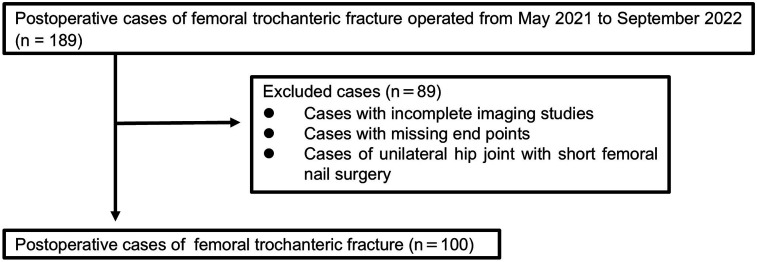
Methods: This multicenter prospective observational study included 100 patients with femoral trochanteric fractures. Preoperative CT images were obtained from the first lumbar vertebra to the distal end of the femur. Using 3D Slicer (version 7), the proximal uninjured femur was segmented and reconstructed into a 3D model. The volume and CT values (Hounsfield units [HU]) of the proximal femur and femoral head were measured. Additionally, CT values were used to assess the bone volume and distribution of low bone-density areas (0-100 HU) and high bone-density areas (≥300 HU) in the femoral head.
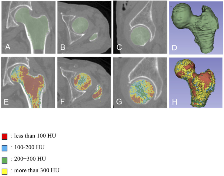
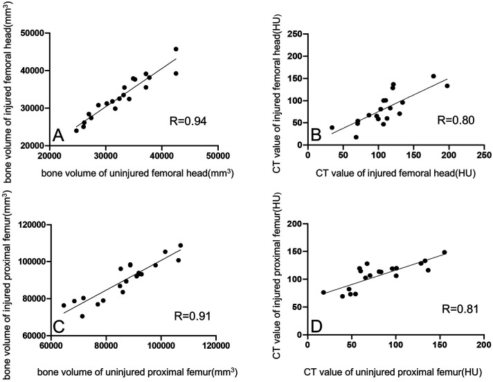
Results: The average bone volume and CT values of proximal femur and femoral head were 90,641 mm3 and 94 HU, and 32,316 mm3 and 131 HU, respectively. The volume of the femoral head with CT values ≥300 HU was 2967 mm3, accounting for 9.1% of the total volume (range: 0.1%-32.6%), with a distribution observed along the central region of the femoral head, particularly along the principal compressive trabeculae. Additionally, the average CT value of the femoral head (<100 HU, ≥300 HU) correlated with bone volume.
Conclusion: A distribution of CT values within the femoral head is characteristic of patients with femoral trochanteric fractures. Using HU values from CT imaging to predict bone fragility preoperatively may aid in assessing the risk of postoperative complications.
期刊介绍:
Geriatric Orthopaedic Surgery & Rehabilitation (GOS) is an open access, peer-reviewed journal that provides clinical information concerning musculoskeletal conditions affecting the aging population. GOS focuses on care of geriatric orthopaedic patients and their subsequent rehabilitation. This journal is a member of the Committee on Publication Ethics (COPE).
 求助内容:
求助内容: 应助结果提醒方式:


