{"title":"Knotting of a Urinary Catheter and Ureteric Stent: A Unique Complication and Management Solution.","authors":"H Logan, K Lockhart, P Chong","doi":"10.1155/criu/5559138","DOIUrl":null,"url":null,"abstract":"<p><p><b>Objective:</b> Spontaneous intravesical knotting is a highly infrequent complication of urinary catheters. We present a novel endoscopic treatment approach to managing a spontaneously knotted urinary catheter around a ureteric stent. <b>Subject:</b> A 79-year-old man presented to the Emergency department with confusion and acute renal failure. His background was significant for metastatic castrate-resistant prostate cancer. His associated obstructive uropathy was managed with a long-term right 7-Fr Rüsch ureteric stent, last changed 1 month prior and a long-term 18-Fr indwelling catheter. A CT intravenous pyelogram clearly demonstrated his indwelling catheter knotted around and through the distal intravesical portion of an appropriately positioned right ureteric stent. <b>Results:</b> Following decompression of the left kidney via percutaneous nephrostomy, attempts were made to remove the urinary catheter under fluoroscopy with a variety of wires and introducers. The patient then underwent a general anesthesia, and the knot was successfully removed piecemeal with a Mauermayer stone crusher via 25-Fr access sheath. <b>Conclusion:</b> Endoscopic techniques such as the use of a stone crusher may be beneficial for the removal of difficult and complex catheter knots as demonstrated in this case. Catheter knotting should always be considered if the functioning or attempted removal of the catheter is abnormal and timely referral to a urologist is made.</p>","PeriodicalId":30323,"journal":{"name":"Case Reports in Urology","volume":"2025 ","pages":"5559138"},"PeriodicalIF":0.0000,"publicationDate":"2025-07-10","publicationTypes":"Journal Article","fieldsOfStudy":null,"isOpenAccess":false,"openAccessPdf":"https://www.ncbi.nlm.nih.gov/pmc/articles/PMC12271699/pdf/","citationCount":"0","resultStr":null,"platform":"Semanticscholar","paperid":null,"PeriodicalName":"Case Reports in Urology","FirstCategoryId":"1085","ListUrlMain":"https://doi.org/10.1155/criu/5559138","RegionNum":0,"RegionCategory":null,"ArticlePicture":[],"TitleCN":null,"AbstractTextCN":null,"PMCID":null,"EPubDate":"2025/1/1 0:00:00","PubModel":"eCollection","JCR":"","JCRName":"","Score":null,"Total":0}
引用次数: 0
Abstract
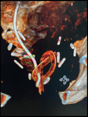
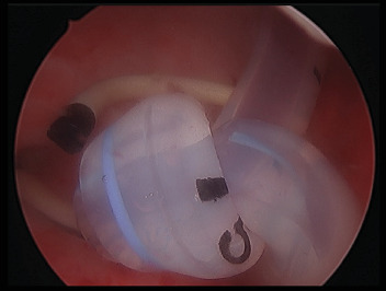
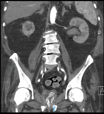
Objective: Spontaneous intravesical knotting is a highly infrequent complication of urinary catheters. We present a novel endoscopic treatment approach to managing a spontaneously knotted urinary catheter around a ureteric stent. Subject: A 79-year-old man presented to the Emergency department with confusion and acute renal failure. His background was significant for metastatic castrate-resistant prostate cancer. His associated obstructive uropathy was managed with a long-term right 7-Fr Rüsch ureteric stent, last changed 1 month prior and a long-term 18-Fr indwelling catheter. A CT intravenous pyelogram clearly demonstrated his indwelling catheter knotted around and through the distal intravesical portion of an appropriately positioned right ureteric stent. Results: Following decompression of the left kidney via percutaneous nephrostomy, attempts were made to remove the urinary catheter under fluoroscopy with a variety of wires and introducers. The patient then underwent a general anesthesia, and the knot was successfully removed piecemeal with a Mauermayer stone crusher via 25-Fr access sheath. Conclusion: Endoscopic techniques such as the use of a stone crusher may be beneficial for the removal of difficult and complex catheter knots as demonstrated in this case. Catheter knotting should always be considered if the functioning or attempted removal of the catheter is abnormal and timely referral to a urologist is made.

 求助内容:
求助内容: 应助结果提醒方式:


