Negar Firoozeh, Spencer C Behr, Antonio C Westphalen
{"title":"Predicting uninformative prostate magnetic resonance imaging sequences: a hypothesis-generating pilot study.","authors":"Negar Firoozeh, Spencer C Behr, Antonio C Westphalen","doi":"10.1590/0100-3984.2025.0007","DOIUrl":null,"url":null,"abstract":"<p><strong>Objective: </strong>To determine the proportion of men with completely negative multiparametric magnetic resonance imaging (MRI) scans and which individual sequence-T2-weighted imaging (T2WI) or diffusion-weighted imaging (DWI)-best predicts an overall negative examination result.</p><p><strong>Materials and methods: </strong>This was a single-center retrospective study evaluating 492 MRI scans compliant with Prostate Imaging Reporting and Data System (PI-RADS), version 2.1. Radiology reports described the absence of lesions or suspicious lesions with PI-RADS scores of 3-5, signifying positive T2WI or DWI results. Positivity on a dynamic contrast-enhanced (DCE) study was determined by early or simultaneous focal enhancement consistent with lesions on T2WI or DWI. All scans reported as negative were prospectively reviewed to ensure that each sequence truly met the criteria for negativity according to the PI-RADS guidelines. Descriptive statistics were employed to summarize the data, and the chi-square test was employed to assess the relationship between a negative T2WI result and a negative DWI/DCE result, as well as that between a negative DWI result and a negative DWI/DCE result, with logistic regression models identifying predictors of such combined results.</p><p><strong>Results: </strong>Among the patients evaluated, approximately one-third of those with suspected prostate cancer and 10% of those with known cancer could have concluded their examination after a single negative sequence. A negative T2WI result predicted negative DWI/DCE findings in 62.4% of scans (95% CI: 55.3-68.9), with an odds ratio of 245.3 (<i>p</i> < 0.001). Similarly, a negative DWI result predicted negative T2WI/DCE findings in 88.9% of scans (95% CI: 83.1-92.7) with an odds ratio of 76.4 (<i>p</i> < 0.001). These associations remained robust after adjustment for age, prostate-specific antigen level, prostate-specific antigen density, cancer status, and radiologist.</p><p><strong>Conclusion: </strong>Findings from T2WI or DWI may serve as preliminary indicators for the subsequent diagnostic yield of other sequences, with DWI appearing to hold a slight advantage. Although the accuracy of this approach is not yet sufficient for clinical implementation, these results are promising and merit further investigation.</p>","PeriodicalId":20842,"journal":{"name":"Radiologia Brasileira","volume":"58 ","pages":"e20250007"},"PeriodicalIF":0.0000,"publicationDate":"2025-07-17","publicationTypes":"Journal Article","fieldsOfStudy":null,"isOpenAccess":false,"openAccessPdf":"https://www.ncbi.nlm.nih.gov/pmc/articles/PMC12274023/pdf/","citationCount":"0","resultStr":null,"platform":"Semanticscholar","paperid":null,"PeriodicalName":"Radiologia Brasileira","FirstCategoryId":"1085","ListUrlMain":"https://doi.org/10.1590/0100-3984.2025.0007","RegionNum":0,"RegionCategory":null,"ArticlePicture":[],"TitleCN":null,"AbstractTextCN":null,"PMCID":null,"EPubDate":"2025/1/1 0:00:00","PubModel":"eCollection","JCR":"Q3","JCRName":"Medicine","Score":null,"Total":0}
引用次数: 0
Abstract
Objective: To determine the proportion of men with completely negative multiparametric magnetic resonance imaging (MRI) scans and which individual sequence-T2-weighted imaging (T2WI) or diffusion-weighted imaging (DWI)-best predicts an overall negative examination result.
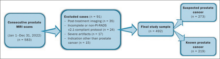
Materials and methods: This was a single-center retrospective study evaluating 492 MRI scans compliant with Prostate Imaging Reporting and Data System (PI-RADS), version 2.1. Radiology reports described the absence of lesions or suspicious lesions with PI-RADS scores of 3-5, signifying positive T2WI or DWI results. Positivity on a dynamic contrast-enhanced (DCE) study was determined by early or simultaneous focal enhancement consistent with lesions on T2WI or DWI. All scans reported as negative were prospectively reviewed to ensure that each sequence truly met the criteria for negativity according to the PI-RADS guidelines. Descriptive statistics were employed to summarize the data, and the chi-square test was employed to assess the relationship between a negative T2WI result and a negative DWI/DCE result, as well as that between a negative DWI result and a negative DWI/DCE result, with logistic regression models identifying predictors of such combined results.
Results: Among the patients evaluated, approximately one-third of those with suspected prostate cancer and 10% of those with known cancer could have concluded their examination after a single negative sequence. A negative T2WI result predicted negative DWI/DCE findings in 62.4% of scans (95% CI: 55.3-68.9), with an odds ratio of 245.3 (p < 0.001). Similarly, a negative DWI result predicted negative T2WI/DCE findings in 88.9% of scans (95% CI: 83.1-92.7) with an odds ratio of 76.4 (p < 0.001). These associations remained robust after adjustment for age, prostate-specific antigen level, prostate-specific antigen density, cancer status, and radiologist.
Conclusion: Findings from T2WI or DWI may serve as preliminary indicators for the subsequent diagnostic yield of other sequences, with DWI appearing to hold a slight advantage. Although the accuracy of this approach is not yet sufficient for clinical implementation, these results are promising and merit further investigation.

 求助内容:
求助内容: 应助结果提醒方式:


