{"title":"Changes in Major Retinal Blood Vessel Position Outside the Optic Nerve Head in Glaucomatous Eyes.","authors":"Zahra Karjou, Shahin Yazdani, Behrouz Alizadeh Savareh, Bahareh Kheiri, Fatemeh Radinmehr","doi":"10.18502/jovr.v20.15461","DOIUrl":null,"url":null,"abstract":"<p><strong>Purpose: </strong>Vascular changes along with loss of the neural rim at the optic nerve head (ONH) are established hallmarks of glaucomatous optic neuropathy. The current study investigates changes in the position of major retinal vessels outside the ONH in eyes with definite or suspected glaucoma and reports its clinical associations.</p><p><strong>Methods: </strong>This retrospective case-control study was conducted on a dataset of 2390 patients with definite or suspected glaucoma and serial photographic documentation from 2015 to 2022. Images were overlaid chronologically and examined for vascular displacement (VD) outside the margin of the ONH up to one disc diameter using the built-in fundus camera software; in the case of VD detection, the change was verified using MATLAB software. The amount of VD was measured in pixels and expressed in a unitless arbitrary ratio derived from the amount of VD in pixels divided by the largest optic disc diameter in pixels. During the study period, a small number of eyes showed evidence of VD, which made up the case group; eyes without evidence of VD from the same dataset were chosen as controls.</p><p><strong>Results: </strong>A total of 23 eyes demonstrated VD, and 60 eyes with no evidence of VD were selected as controls. The mean amount of VD was 0.15 <math><mo>±</mo></math> 0.01 in case eyes compared to 0.01 <math><mo>±</mo></math> 0.01 in control eyes (<i>P</i> <math><mo><</mo></math> 0.001). Definite glaucomatous damage was observed in 20 (87%) eyes in the case group compared to 35 (58.3%) eyes in the control group (<i>P</i> = 0.014). The best-corrected visual acuity in eyes with VD, both at baseline and at the final visit, was significantly worse than in controls (<i>P</i> = 0.018 and <i>P</i> = 0.032, respectively). Eyes with VD had greater cupping both at baseline (<i>P</i> = 0.025) and at the final examination (<i>P</i> = 0.04). During the study period, 16 (69.6%) eyes with VD and 12 (20%) control eyes required glaucoma surgery (<i>P</i> = 0.001). Patients with VD also showed a statistical trend toward being younger (mean age, 54.5 <math><mo>±</mo></math> 16.5 vs 61.3 <math><mo>±</mo></math> 15.5 years, <i>P</i> = 0.088).</p><p><strong>Conclusion: </strong>VD outside the ONH may occur in eyes with glaucoma and is associated with factors reflecting more significant glaucomatous damage. Eyes with VD outside the ONH have lower visual acuity, greater cupping, and require glaucoma surgery more often, indicating more significant glaucoma severity or progression.</p>","PeriodicalId":16586,"journal":{"name":"Journal of Ophthalmic & Vision Research","volume":"20 ","pages":""},"PeriodicalIF":1.5000,"publicationDate":"2025-07-10","publicationTypes":"Journal Article","fieldsOfStudy":null,"isOpenAccess":false,"openAccessPdf":"https://www.ncbi.nlm.nih.gov/pmc/articles/PMC12261259/pdf/","citationCount":"0","resultStr":null,"platform":"Semanticscholar","paperid":null,"PeriodicalName":"Journal of Ophthalmic & Vision Research","FirstCategoryId":"1085","ListUrlMain":"https://doi.org/10.18502/jovr.v20.15461","RegionNum":0,"RegionCategory":null,"ArticlePicture":[],"TitleCN":null,"AbstractTextCN":null,"PMCID":null,"EPubDate":"2025/1/1 0:00:00","PubModel":"eCollection","JCR":"Q3","JCRName":"OPHTHALMOLOGY","Score":null,"Total":0}
引用次数: 0
Abstract
Purpose: Vascular changes along with loss of the neural rim at the optic nerve head (ONH) are established hallmarks of glaucomatous optic neuropathy. The current study investigates changes in the position of major retinal vessels outside the ONH in eyes with definite or suspected glaucoma and reports its clinical associations.
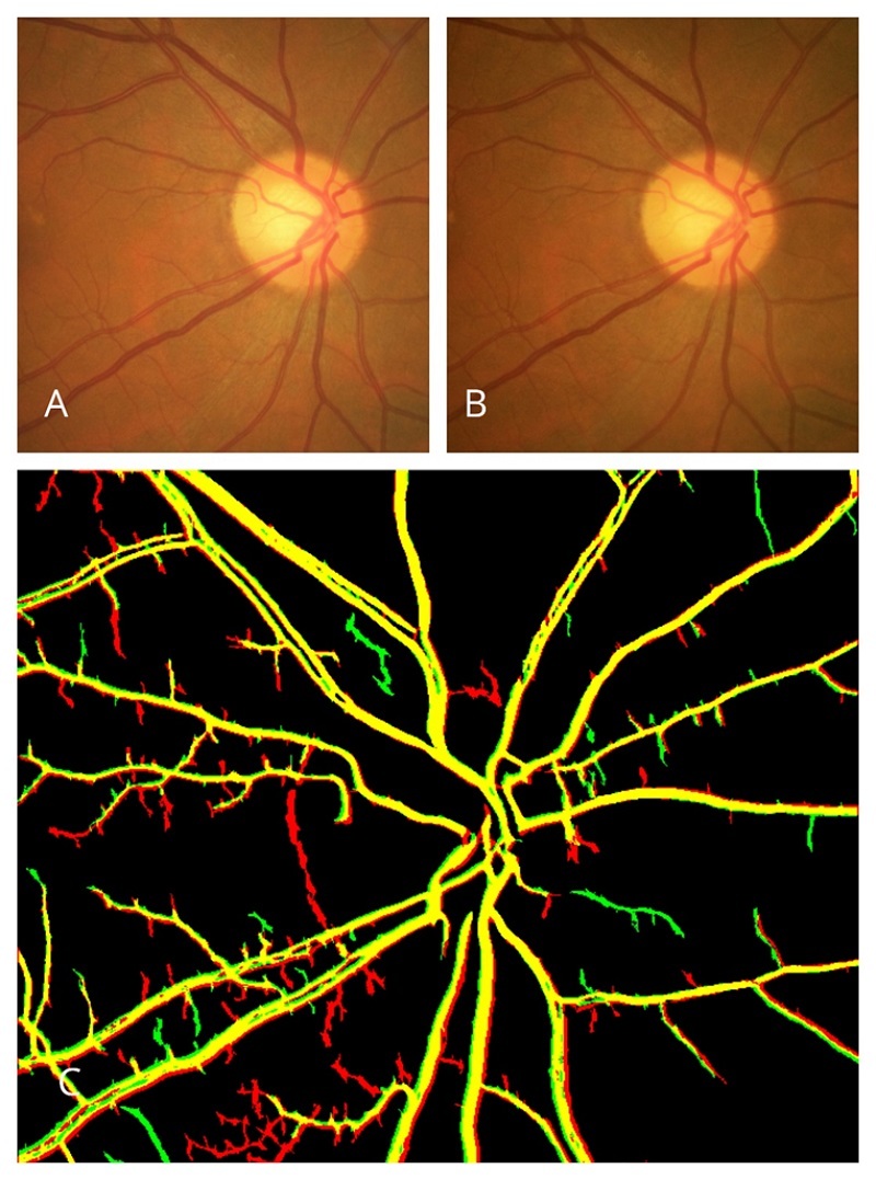
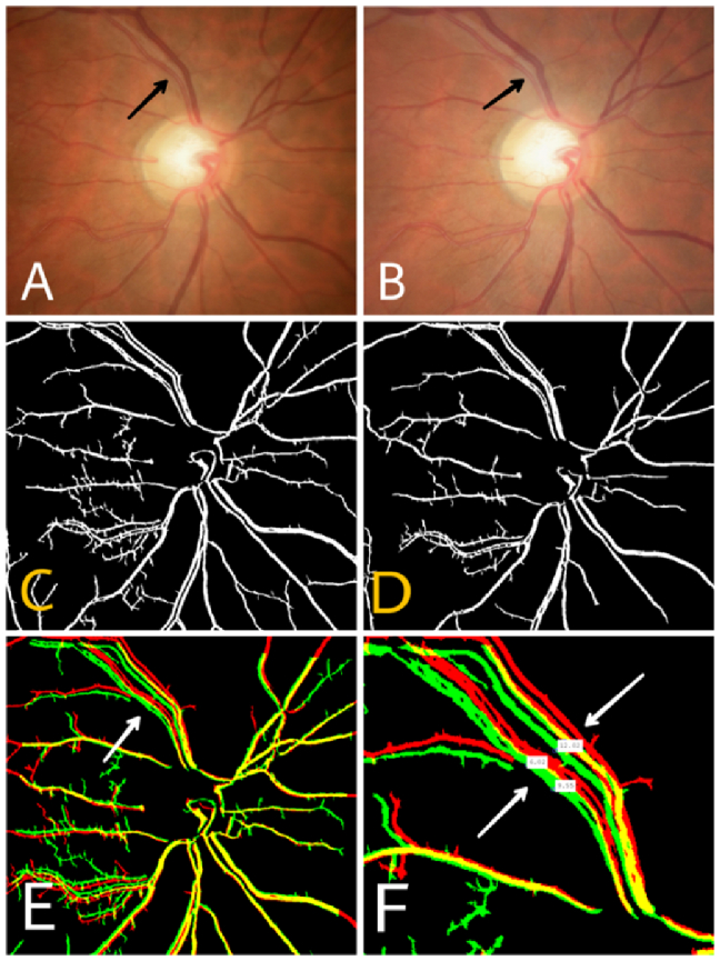
Methods: This retrospective case-control study was conducted on a dataset of 2390 patients with definite or suspected glaucoma and serial photographic documentation from 2015 to 2022. Images were overlaid chronologically and examined for vascular displacement (VD) outside the margin of the ONH up to one disc diameter using the built-in fundus camera software; in the case of VD detection, the change was verified using MATLAB software. The amount of VD was measured in pixels and expressed in a unitless arbitrary ratio derived from the amount of VD in pixels divided by the largest optic disc diameter in pixels. During the study period, a small number of eyes showed evidence of VD, which made up the case group; eyes without evidence of VD from the same dataset were chosen as controls.
Results: A total of 23 eyes demonstrated VD, and 60 eyes with no evidence of VD were selected as controls. The mean amount of VD was 0.15 0.01 in case eyes compared to 0.01 0.01 in control eyes (P 0.001). Definite glaucomatous damage was observed in 20 (87%) eyes in the case group compared to 35 (58.3%) eyes in the control group (P = 0.014). The best-corrected visual acuity in eyes with VD, both at baseline and at the final visit, was significantly worse than in controls (P = 0.018 and P = 0.032, respectively). Eyes with VD had greater cupping both at baseline (P = 0.025) and at the final examination (P = 0.04). During the study period, 16 (69.6%) eyes with VD and 12 (20%) control eyes required glaucoma surgery (P = 0.001). Patients with VD also showed a statistical trend toward being younger (mean age, 54.5 16.5 vs 61.3 15.5 years, P = 0.088).
Conclusion: VD outside the ONH may occur in eyes with glaucoma and is associated with factors reflecting more significant glaucomatous damage. Eyes with VD outside the ONH have lower visual acuity, greater cupping, and require glaucoma surgery more often, indicating more significant glaucoma severity or progression.

 求助内容:
求助内容: 应助结果提醒方式:


