Differential expression of haematopoietic prostaglandin D synthase by POU2F3‑positive tuft cells in conventional bilayered oncocytic and metaplastic epithelia of Warthin tumours.
{"title":"Differential expression of haematopoietic prostaglandin D synthase by POU2F3‑positive tuft cells in conventional bilayered oncocytic and metaplastic epithelia of Warthin tumours.","authors":"Kenta Hosomi, Akira Sato, Mitsuaki Ishida, Kensuke Nakanishi, Tetsuya Terada, Shin-Ichi Haginomori, Yoshinobu Hirose, Ko Fujimori","doi":"10.3892/mmr.2025.13624","DOIUrl":null,"url":null,"abstract":"<p><p>Warthin tumours (WT), the second most common benign salivary gland tumour, are histopathologically composed of bilayered oncocytic epithelial cells with occasional metaplastic epithelium. Tuft cells, which are chemosensory epithelial cells, are present in WT. Tuft cells serve various roles by producing physiologically active substances, such as prostaglandins (PGs). PGD2 released from tuft cells is crucial for tissue repair and inhibiting pancreatic carcinogenesis. However, whether or not tuft cells in WT produce PGD2 has not yet been elucidated. The present study aimed to investigate the production of PGD<sub>2</sub> in POU class 2 homeobox 3 (POU2F3; a specific tuft cell marker)‑positive cells of WT and normal salivary glands. Consecutive patients with WT who underwent surgical resection were selected. Dual immunohistochemical staining for POU2F3 and haematopoietic PGD synthase (H‑PGDS) was performed. The present study included 28 patients with WT of the parotid gland (all male patients; median age, 68 years). The conventional bilayered oncocytic epithelium was present in all tumours; squamous metaplastic epithelium and conventional bilayered oncocytic epithelium were observed in 16 patients. Dual immunohistochemical analysis revealed that POU2F3+/H‑PGDS‑ cells were exclusively present in the striated duct of the normal salivary gland, and abundant POU2F3‑positive tuft cells were observed in both the conventional bilayered oncocytic and metaplastic squamous epithelia of WT. The median ratio of POU2F3‑positive cells expressing H‑PGDS was significantly higher in the conventional oncocytic epithelium (89.9%) than in the metaplastic squamous epithelium (10.6%) of WT (P=0.00044). The present results suggest a link between tissue injury to the striated duct and the pathogenesis of WT, and that PGD<sub>2</sub> released from POU2F3‑positive cells in the conventional bilayered oncocytic epithelium is associated with ongoing tissue injury. Further studies are warranted to clarify the function of tuft cells in WT and gain deeper insights into the pathogenesis of WT.</p>","PeriodicalId":18818,"journal":{"name":"Molecular medicine reports","volume":"32 4","pages":""},"PeriodicalIF":3.5000,"publicationDate":"2025-10-01","publicationTypes":"Journal Article","fieldsOfStudy":null,"isOpenAccess":false,"openAccessPdf":"https://www.ncbi.nlm.nih.gov/pmc/articles/PMC12308854/pdf/","citationCount":"0","resultStr":null,"platform":"Semanticscholar","paperid":null,"PeriodicalName":"Molecular medicine reports","FirstCategoryId":"3","ListUrlMain":"https://doi.org/10.3892/mmr.2025.13624","RegionNum":3,"RegionCategory":"医学","ArticlePicture":[],"TitleCN":null,"AbstractTextCN":null,"PMCID":null,"EPubDate":"2025/7/19 0:00:00","PubModel":"Epub","JCR":"Q2","JCRName":"MEDICINE, RESEARCH & EXPERIMENTAL","Score":null,"Total":0}
引用次数: 0
Abstract
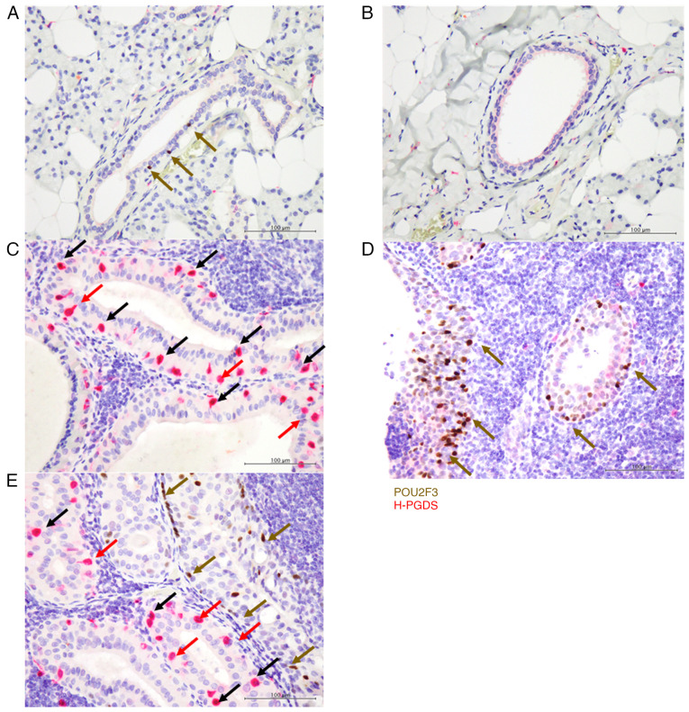
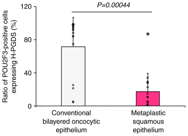
Warthin tumours (WT), the second most common benign salivary gland tumour, are histopathologically composed of bilayered oncocytic epithelial cells with occasional metaplastic epithelium. Tuft cells, which are chemosensory epithelial cells, are present in WT. Tuft cells serve various roles by producing physiologically active substances, such as prostaglandins (PGs). PGD2 released from tuft cells is crucial for tissue repair and inhibiting pancreatic carcinogenesis. However, whether or not tuft cells in WT produce PGD2 has not yet been elucidated. The present study aimed to investigate the production of PGD2 in POU class 2 homeobox 3 (POU2F3; a specific tuft cell marker)‑positive cells of WT and normal salivary glands. Consecutive patients with WT who underwent surgical resection were selected. Dual immunohistochemical staining for POU2F3 and haematopoietic PGD synthase (H‑PGDS) was performed. The present study included 28 patients with WT of the parotid gland (all male patients; median age, 68 years). The conventional bilayered oncocytic epithelium was present in all tumours; squamous metaplastic epithelium and conventional bilayered oncocytic epithelium were observed in 16 patients. Dual immunohistochemical analysis revealed that POU2F3+/H‑PGDS‑ cells were exclusively present in the striated duct of the normal salivary gland, and abundant POU2F3‑positive tuft cells were observed in both the conventional bilayered oncocytic and metaplastic squamous epithelia of WT. The median ratio of POU2F3‑positive cells expressing H‑PGDS was significantly higher in the conventional oncocytic epithelium (89.9%) than in the metaplastic squamous epithelium (10.6%) of WT (P=0.00044). The present results suggest a link between tissue injury to the striated duct and the pathogenesis of WT, and that PGD2 released from POU2F3‑positive cells in the conventional bilayered oncocytic epithelium is associated with ongoing tissue injury. Further studies are warranted to clarify the function of tuft cells in WT and gain deeper insights into the pathogenesis of WT.
期刊介绍:
Molecular Medicine Reports is a monthly, peer-reviewed journal available in print and online, that includes studies devoted to molecular medicine, underscoring aspects including pharmacology, pathology, genetics, neurosciences, infectious diseases, molecular cardiology and molecular surgery. In vitro and in vivo studies of experimental model systems pertaining to the mechanisms of a variety of diseases offer researchers the necessary tools and knowledge with which to aid the diagnosis and treatment of human diseases.
 求助内容:
求助内容: 应助结果提醒方式:


