{"title":"Using Convoluted Neural Networks in Diagnosing Lung Cancer on Computed Tomography Scans.","authors":"Ovidiu Cîmpeanu, Ilona Mihaela Liliac, Mădălin Mămuleanu, Ștefan-Vlad Voinea, Mihai Olteanu, Costin-Teodor Streba","doi":"10.12865/CHSJ.51.01.09","DOIUrl":null,"url":null,"abstract":"<p><strong>Introduction: </strong>Lung cancer represents a major health issue of the modern world, accounting for both most new cases and highest mortality rates worldwide. Early diagnosis and treatment remain essential in managing the disease; therefore, developing novel computer-assisted tools for processing large quantities of imaging data can prove indispensable. Our aim was to develop a novel convoluted neural network (CNN) to classify lung computed tomography (CT) images of suspect nodules.</p><p><strong>Materials and methods: </strong>After obtaining ethical clearance, we included consenting patients with a lung mass found on a chest radiography, visible lung tumor on computer tomography and positive pathology or follow-up. After data augmentation, we trained a deep learning model to classify input images into two classes, malignant or benign. We evaluated the model by calculating accuracy, recall and precision.</p><p><strong>Results: </strong>We successfully enrolled 176 patients from a total of 192 cases. Most were male (135 cases, accounting for 76.7%) and came from urban areas (111 cases, 63%). Most tumors were found on the right lung (103 cases). The model performed well on an imbalanced dataset, with recall values at 79.31%, while precision reached 62.16%, a training accuracy of 76.34% and a validation accuracy of 77.01%.</p><p><strong>Conclusions: </strong>We proved that a CNN model can easily be implemented on regular hardware to successfully classify malignant and benign lung lesions on CT images. Future CNN implementations can greatly improve the imaging diagnosis of lung lesions; however, the physicians should always decide the medical management.</p>","PeriodicalId":93963,"journal":{"name":"Current health sciences journal","volume":"51 1","pages":"87-95"},"PeriodicalIF":0.0000,"publicationDate":"2025-01-01","publicationTypes":"Journal Article","fieldsOfStudy":null,"isOpenAccess":false,"openAccessPdf":"https://www.ncbi.nlm.nih.gov/pmc/articles/PMC12264995/pdf/","citationCount":"0","resultStr":null,"platform":"Semanticscholar","paperid":null,"PeriodicalName":"Current health sciences journal","FirstCategoryId":"1085","ListUrlMain":"https://doi.org/10.12865/CHSJ.51.01.09","RegionNum":0,"RegionCategory":null,"ArticlePicture":[],"TitleCN":null,"AbstractTextCN":null,"PMCID":null,"EPubDate":"2025/3/31 0:00:00","PubModel":"Epub","JCR":"","JCRName":"","Score":null,"Total":0}
引用次数: 0
Abstract
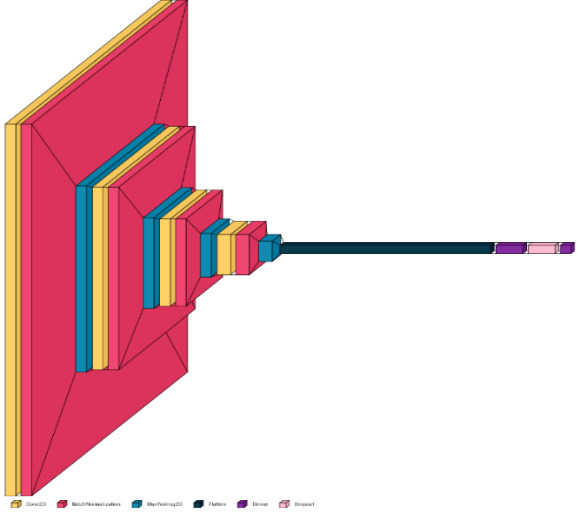
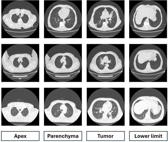
Introduction: Lung cancer represents a major health issue of the modern world, accounting for both most new cases and highest mortality rates worldwide. Early diagnosis and treatment remain essential in managing the disease; therefore, developing novel computer-assisted tools for processing large quantities of imaging data can prove indispensable. Our aim was to develop a novel convoluted neural network (CNN) to classify lung computed tomography (CT) images of suspect nodules.
Materials and methods: After obtaining ethical clearance, we included consenting patients with a lung mass found on a chest radiography, visible lung tumor on computer tomography and positive pathology or follow-up. After data augmentation, we trained a deep learning model to classify input images into two classes, malignant or benign. We evaluated the model by calculating accuracy, recall and precision.
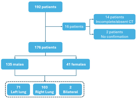
Results: We successfully enrolled 176 patients from a total of 192 cases. Most were male (135 cases, accounting for 76.7%) and came from urban areas (111 cases, 63%). Most tumors were found on the right lung (103 cases). The model performed well on an imbalanced dataset, with recall values at 79.31%, while precision reached 62.16%, a training accuracy of 76.34% and a validation accuracy of 77.01%.
Conclusions: We proved that a CNN model can easily be implemented on regular hardware to successfully classify malignant and benign lung lesions on CT images. Future CNN implementations can greatly improve the imaging diagnosis of lung lesions; however, the physicians should always decide the medical management.

 求助内容:
求助内容: 应助结果提醒方式:


