{"title":"Tertiary Dentin Barrier Formation: A Comparison Between the Effects of Two Calcium Silicate Based Materials.","authors":"Antoanela Magdalena Covaci, Mihai Andrei, Ioana Dinca, Lucian Toma Ciocan, Madalina Nicoleta Matei, Andreea Cristiana Didilescu","doi":"10.12865/CHSJ.51.01.10","DOIUrl":null,"url":null,"abstract":"<p><p>Pulp capping is a vital pulp therapy that aims to prolong the life of a tooth by protecting it after pulp exposure occurred. Pulp capping biomaterials are intended to induce odontoblasts to deposit a natural tertiary dentin barrier to protect the pulp-dentin complex. Two pulp capping agents with calcium silicates in their composition, but with different mechanisms of the setting reaction were tested in vivo with the main objective of comparing their effect on the pulp-dentin complex. The specific aim was to evaluate the preservation of pulp vitality following in vivo direct and indirect pulp capping on eight human third molars. TheraCal LC, a light-cured calcium silicate-based material, was tested both by direct and indirect pulp capping, while the mineral trioxide aggregate (MTA) cement was tested by direct pulp capping. The molars were assessed by micro-computed tomography (micro-CT) and by light microscopy and stereo-microscopy following histological processing of the teeth. Dental pulp vitality testing was performed before tooth extraction. Inflammatory pulp status was performed on light microscopy images and it was investigated the presence of inflammatory infiltrate, edema, vascular congestion and pulp necrosis. Following pulp capping, the MTA cement showed more favorable results, generating the formation of complete or incomplete dentin bridges in all treated teeth, while TheraCal LC induced the formation of dentin bridges in only two teeth. Tooth vitality was preserved in all tested teeth. In conclusion, both materials stimulated neodentinogenesis, with the MTA cement being more effective and presenting a much more favorable biological pulpal response.</p>","PeriodicalId":93963,"journal":{"name":"Current health sciences journal","volume":"51 1","pages":"96-104"},"PeriodicalIF":0.0000,"publicationDate":"2025-01-01","publicationTypes":"Journal Article","fieldsOfStudy":null,"isOpenAccess":false,"openAccessPdf":"https://www.ncbi.nlm.nih.gov/pmc/articles/PMC12264994/pdf/","citationCount":"0","resultStr":null,"platform":"Semanticscholar","paperid":null,"PeriodicalName":"Current health sciences journal","FirstCategoryId":"1085","ListUrlMain":"https://doi.org/10.12865/CHSJ.51.01.10","RegionNum":0,"RegionCategory":null,"ArticlePicture":[],"TitleCN":null,"AbstractTextCN":null,"PMCID":null,"EPubDate":"2025/3/31 0:00:00","PubModel":"Epub","JCR":"","JCRName":"","Score":null,"Total":0}
引用次数: 0
Abstract
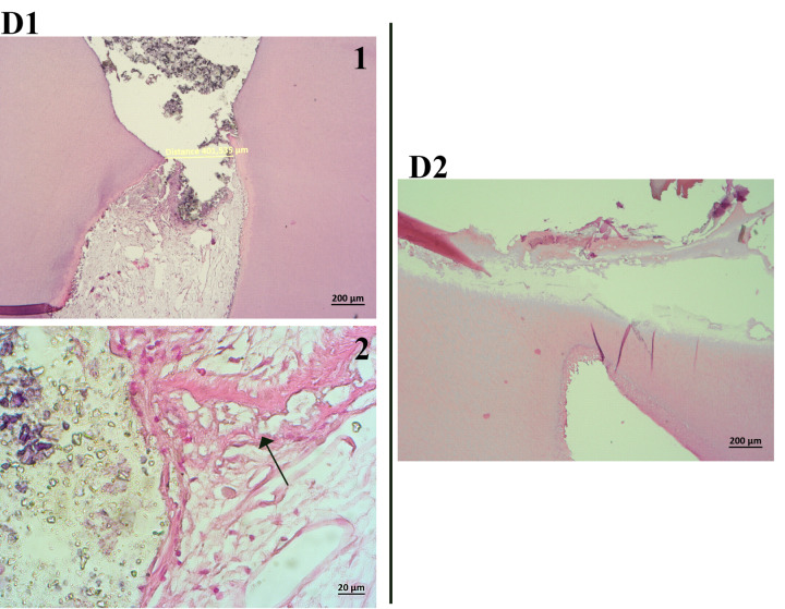
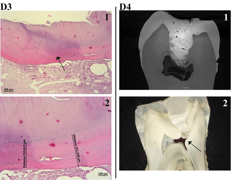
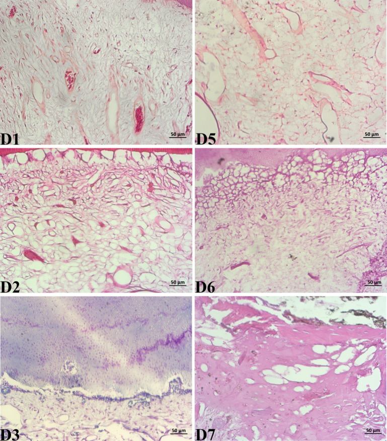
Pulp capping is a vital pulp therapy that aims to prolong the life of a tooth by protecting it after pulp exposure occurred. Pulp capping biomaterials are intended to induce odontoblasts to deposit a natural tertiary dentin barrier to protect the pulp-dentin complex. Two pulp capping agents with calcium silicates in their composition, but with different mechanisms of the setting reaction were tested in vivo with the main objective of comparing their effect on the pulp-dentin complex. The specific aim was to evaluate the preservation of pulp vitality following in vivo direct and indirect pulp capping on eight human third molars. TheraCal LC, a light-cured calcium silicate-based material, was tested both by direct and indirect pulp capping, while the mineral trioxide aggregate (MTA) cement was tested by direct pulp capping. The molars were assessed by micro-computed tomography (micro-CT) and by light microscopy and stereo-microscopy following histological processing of the teeth. Dental pulp vitality testing was performed before tooth extraction. Inflammatory pulp status was performed on light microscopy images and it was investigated the presence of inflammatory infiltrate, edema, vascular congestion and pulp necrosis. Following pulp capping, the MTA cement showed more favorable results, generating the formation of complete or incomplete dentin bridges in all treated teeth, while TheraCal LC induced the formation of dentin bridges in only two teeth. Tooth vitality was preserved in all tested teeth. In conclusion, both materials stimulated neodentinogenesis, with the MTA cement being more effective and presenting a much more favorable biological pulpal response.

 求助内容:
求助内容: 应助结果提醒方式:


