Gergely David, Alice Motovylyak, Felix Schlegel, Zsofia Kovacs, Christian Kündig, Angela R Filous, Jan M Schwab, Matthew D Budde, Jan Klohs, Patrick Freund
{"title":"Progressive Remote Axonal Degeneration Following Spinal Cord Injury: A Histological and MRI Study.","authors":"Gergely David, Alice Motovylyak, Felix Schlegel, Zsofia Kovacs, Christian Kündig, Angela R Filous, Jan M Schwab, Matthew D Budde, Jan Klohs, Patrick Freund","doi":"10.1089/neur.2025.0011","DOIUrl":null,"url":null,"abstract":"<p><p>In acute human spinal cord injury (SCI), magnetic resonance imaging (MRI) reveals progressive neuroanatomical changes at the lesion site and in remote regions. Here, we aimed to elucidate the structural underpinnings of these neuroanatomical changes and to characterize their spatiotemporal distribution in a rat contusion SCI model, using both histology and MRI. First, rats subjected to a thoracic contusion SCI (T8) and sham-operated rats were sacrificed at 56 days post-injury (dpi), and SMI-32 immunohistochemistry was used to assess remote axonal degeneration at cervical segments C2-C5. Second, to evaluate the effect of severity and time since injury on axonal degeneration, rats of varying injury severity were sacrificed at 2, 30, and 90 dpi, respectively, followed by SMI-32 immunohistochemistry. Third, <i>ex vivo</i> structural MRI and diffusion tensor imaging were performed rostral to the injury site (C3-T6) at 90 dpi. Histological evidence of axonal degeneration emerged as early as 2 dpi rostral to the injury site, persisting at 90 dpi. Severity-dependent degeneration occurred within the fasciculus gracilis and the periphery of the medio- and ventrolateral columns. Corresponding MRI changes, including lower fractional anisotropy in these regions and smaller gray matter area, were detected. In contrast, the dorsal corticospinal tract exhibited lower fractional anisotropy without clear histological abnormalities, potentially due to atrophy-related mislocalization. This highlights the value of correlative, multimodal approaches and the need for further methodological refinement. The number of SMI-32+ axonal profiles correlated negatively, while gray matter area and fractional anisotropy correlated positively with locomotion assessed by Basso, Beattie, and Bresnahan scores. This study demonstrates in independent experiments that neuroanatomical MRI changes observed after SCI, occurring remote from the injury site, are linked to axonal degeneration. Experimental SCI offers translational insights into underlying mechanisms and potential avenues for neuroprotective or rehabilitative approaches.</p>","PeriodicalId":74300,"journal":{"name":"Neurotrauma reports","volume":"6 1","pages":"443-464"},"PeriodicalIF":1.8000,"publicationDate":"2025-06-05","publicationTypes":"Journal Article","fieldsOfStudy":null,"isOpenAccess":false,"openAccessPdf":"https://www.ncbi.nlm.nih.gov/pmc/articles/PMC12270540/pdf/","citationCount":"0","resultStr":null,"platform":"Semanticscholar","paperid":null,"PeriodicalName":"Neurotrauma reports","FirstCategoryId":"1085","ListUrlMain":"https://doi.org/10.1089/neur.2025.0011","RegionNum":0,"RegionCategory":null,"ArticlePicture":[],"TitleCN":null,"AbstractTextCN":null,"PMCID":null,"EPubDate":"2025/1/1 0:00:00","PubModel":"eCollection","JCR":"Q3","JCRName":"CLINICAL NEUROLOGY","Score":null,"Total":0}
引用次数: 0
Abstract
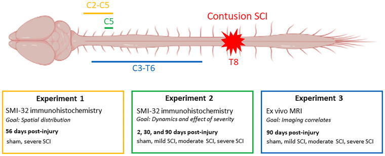
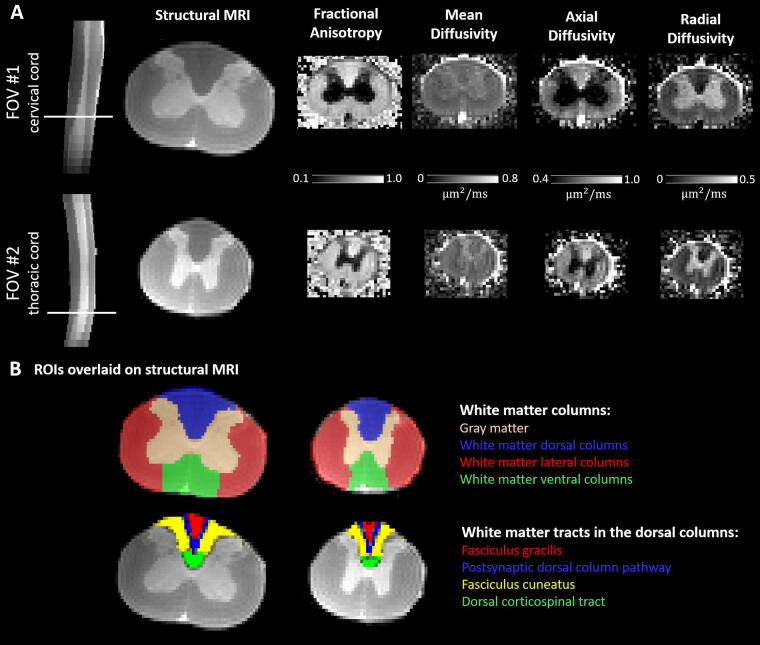
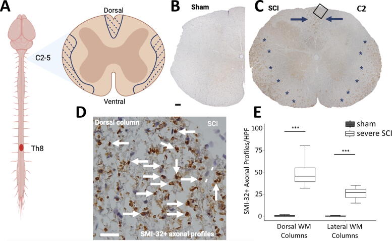
In acute human spinal cord injury (SCI), magnetic resonance imaging (MRI) reveals progressive neuroanatomical changes at the lesion site and in remote regions. Here, we aimed to elucidate the structural underpinnings of these neuroanatomical changes and to characterize their spatiotemporal distribution in a rat contusion SCI model, using both histology and MRI. First, rats subjected to a thoracic contusion SCI (T8) and sham-operated rats were sacrificed at 56 days post-injury (dpi), and SMI-32 immunohistochemistry was used to assess remote axonal degeneration at cervical segments C2-C5. Second, to evaluate the effect of severity and time since injury on axonal degeneration, rats of varying injury severity were sacrificed at 2, 30, and 90 dpi, respectively, followed by SMI-32 immunohistochemistry. Third, ex vivo structural MRI and diffusion tensor imaging were performed rostral to the injury site (C3-T6) at 90 dpi. Histological evidence of axonal degeneration emerged as early as 2 dpi rostral to the injury site, persisting at 90 dpi. Severity-dependent degeneration occurred within the fasciculus gracilis and the periphery of the medio- and ventrolateral columns. Corresponding MRI changes, including lower fractional anisotropy in these regions and smaller gray matter area, were detected. In contrast, the dorsal corticospinal tract exhibited lower fractional anisotropy without clear histological abnormalities, potentially due to atrophy-related mislocalization. This highlights the value of correlative, multimodal approaches and the need for further methodological refinement. The number of SMI-32+ axonal profiles correlated negatively, while gray matter area and fractional anisotropy correlated positively with locomotion assessed by Basso, Beattie, and Bresnahan scores. This study demonstrates in independent experiments that neuroanatomical MRI changes observed after SCI, occurring remote from the injury site, are linked to axonal degeneration. Experimental SCI offers translational insights into underlying mechanisms and potential avenues for neuroprotective or rehabilitative approaches.

 求助内容:
求助内容: 应助结果提醒方式:


