{"title":"A Case of Inflammatory Myofibroblastic Tumor in the Abdominal Wall with Anaplastic Lymphoma Kinase and Whole Exome Sequencing Analysis.","authors":"Yuya Takahata, Shoichi Hazama, Toshiyuki Fujii, Masahiro Kitahara, Keisuke Hino, Kiwamu Okita, Hiroaki Nagano, Ryouichi Tsunedomi, Hiroshi Hashiyada, Kembu Nakamoto","doi":"10.70352/scrj.cr.25-0181","DOIUrl":null,"url":null,"abstract":"<p><strong>Introduction: </strong>Inflammatory myofibroblastic tumors (IMTs) are rare mesenchymal neoplasms characterized by spindle cell proliferation and inflammatory infiltration, but with an unclear etiology. Although IMTs most commonly arise in the lungs, extrapulmonary cases have been documented at various anatomical sites. Approximately 50% of IMTs harbor anaplastic lymphoma kinase (ALK) rearrangements; however, the genetic landscape of ALK-negative cases remains largely unknown. We report a rapidly growing IMT in the right rectus abdominis muscle and present whole-exome sequencing (WES) findings that revealed novel genetic mutations beyond ALK rearrangements.</p><p><strong>Case presentation: </strong>A 38-year-old woman with no significant medical history presented with a rapidly enlarging mass in the right lower abdomen. Computed tomography showed a well-defined tumor on the dorsal side of the right rectus abdominis muscle exhibiting progressive enhancement. Fine-needle biopsy initially suggested the presence of proliferative fasciitis. Owing to rapid tumor growth from 40 to 61 mm within 3 months, laparoscopic surgical resection was performed, including a portion of the posterior sheath and rectus abdominis muscle. Pathological examination confirmed the presence of an IMT and revealed spindle cell proliferation, nuclear atypia, and inflammatory infiltration. Immunohistochemical analysis revealed positivity for smooth muscle actin (SMA) and ALK, partial positivity for desmin, and negativity for cluster of differentiation 34 (CD34) and cytokeratin, compatible with an IMT. WES identified 7 genetic mutations, none of which have been previously reported for IMT in the catalogue of somatic mutations in cancer (COSMIC) database, suggesting novel genetic associations.</p><p><strong>Conclusions: </strong>This case highlights a rare and rapidly growing IMT in the rectus abdominis muscle and underscores the value of molecular analysis in understanding the pathogenesis of IMT. Identification of novel mutations through WES expands the genetic landscape of IMT and may provide insights into tumorigenesis and potential therapeutic targets. Further research is required to explore the clinical implications of these mutations in IMT progression and treatment.</p>","PeriodicalId":22096,"journal":{"name":"Surgical Case Reports","volume":"11 1","pages":""},"PeriodicalIF":0.7000,"publicationDate":"2025-01-01","publicationTypes":"Journal Article","fieldsOfStudy":null,"isOpenAccess":false,"openAccessPdf":"https://www.ncbi.nlm.nih.gov/pmc/articles/PMC12268362/pdf/","citationCount":"0","resultStr":null,"platform":"Semanticscholar","paperid":null,"PeriodicalName":"Surgical Case Reports","FirstCategoryId":"1085","ListUrlMain":"https://doi.org/10.70352/scrj.cr.25-0181","RegionNum":0,"RegionCategory":null,"ArticlePicture":[],"TitleCN":null,"AbstractTextCN":null,"PMCID":null,"EPubDate":"2025/7/16 0:00:00","PubModel":"Epub","JCR":"Q4","JCRName":"SURGERY","Score":null,"Total":0}
引用次数: 0
Abstract
Introduction: Inflammatory myofibroblastic tumors (IMTs) are rare mesenchymal neoplasms characterized by spindle cell proliferation and inflammatory infiltration, but with an unclear etiology. Although IMTs most commonly arise in the lungs, extrapulmonary cases have been documented at various anatomical sites. Approximately 50% of IMTs harbor anaplastic lymphoma kinase (ALK) rearrangements; however, the genetic landscape of ALK-negative cases remains largely unknown. We report a rapidly growing IMT in the right rectus abdominis muscle and present whole-exome sequencing (WES) findings that revealed novel genetic mutations beyond ALK rearrangements.
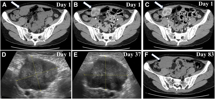
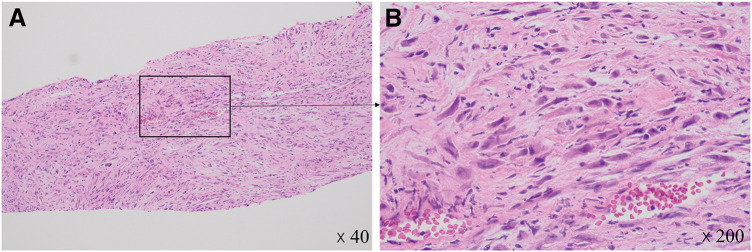
Case presentation: A 38-year-old woman with no significant medical history presented with a rapidly enlarging mass in the right lower abdomen. Computed tomography showed a well-defined tumor on the dorsal side of the right rectus abdominis muscle exhibiting progressive enhancement. Fine-needle biopsy initially suggested the presence of proliferative fasciitis. Owing to rapid tumor growth from 40 to 61 mm within 3 months, laparoscopic surgical resection was performed, including a portion of the posterior sheath and rectus abdominis muscle. Pathological examination confirmed the presence of an IMT and revealed spindle cell proliferation, nuclear atypia, and inflammatory infiltration. Immunohistochemical analysis revealed positivity for smooth muscle actin (SMA) and ALK, partial positivity for desmin, and negativity for cluster of differentiation 34 (CD34) and cytokeratin, compatible with an IMT. WES identified 7 genetic mutations, none of which have been previously reported for IMT in the catalogue of somatic mutations in cancer (COSMIC) database, suggesting novel genetic associations.
Conclusions: This case highlights a rare and rapidly growing IMT in the rectus abdominis muscle and underscores the value of molecular analysis in understanding the pathogenesis of IMT. Identification of novel mutations through WES expands the genetic landscape of IMT and may provide insights into tumorigenesis and potential therapeutic targets. Further research is required to explore the clinical implications of these mutations in IMT progression and treatment.


 求助内容:
求助内容: 应助结果提醒方式:


