{"title":"Power spectrum curve compensation method for image quality enhancement in high-throughput gene sequencing.","authors":"Jianglan Wang, Siyuan Yao, Suocheng Duan, Yu Jiao, Xin Zhang, Xindong Chen","doi":"10.1364/BOE.562958","DOIUrl":null,"url":null,"abstract":"<p><p>In high-throughput gene sequencing, the quality of sequencing images is critical for the accuracy of subsequent base calling. However, during practical sequencing processes, the time delay integration (TDI) camera's push-scan imaging often leads to significant degradation of image quality along the push-scan direction. Addressing the current limitations in TDI image restoration research for gene sequencing, this study establishes an imaging spectrum model of sequencing images based on MGI's ultra-high-throughput sequencer. We systematically analyze the causes and intrinsic mechanisms of image quality degradation, with a focus on elucidating the specific impacts of TDI push-scanning on image quality. To enhance TDI sequencing image quality, we compare the differences in power spectral projection curves between stare-mode imaging and TDI push-scan imaging and propose a power spectrum curve compensation (PSCC)-based quality optimization method alongside a novel evaluation framework for sequencing image quality. Experimental results demonstrate that compared to original H-channel images from cycle 1 to 50, the energy concentration (1/<i>σ</i>) of the optimized images increases by 9.13% in the TDI direction and 4.64% in the direction perpendicular to TDI. Signal-to-noise ratio (SNR) increases by 6.90% for base A and 4.99% for base C, while base calling accuracy (Q30) improves by 1.67%.</p>","PeriodicalId":8969,"journal":{"name":"Biomedical optics express","volume":"16 6","pages":"2351-2364"},"PeriodicalIF":3.2000,"publicationDate":"2025-05-15","publicationTypes":"Journal Article","fieldsOfStudy":null,"isOpenAccess":false,"openAccessPdf":"https://www.ncbi.nlm.nih.gov/pmc/articles/PMC12265594/pdf/","citationCount":"0","resultStr":null,"platform":"Semanticscholar","paperid":null,"PeriodicalName":"Biomedical optics express","FirstCategoryId":"3","ListUrlMain":"https://doi.org/10.1364/BOE.562958","RegionNum":2,"RegionCategory":"医学","ArticlePicture":[],"TitleCN":null,"AbstractTextCN":null,"PMCID":null,"EPubDate":"2025/6/1 0:00:00","PubModel":"eCollection","JCR":"Q2","JCRName":"BIOCHEMICAL RESEARCH METHODS","Score":null,"Total":0}
引用次数: 0
Abstract
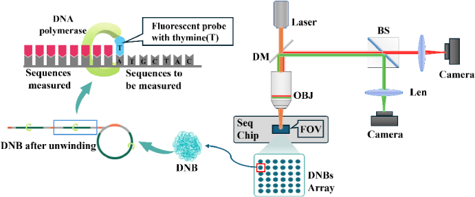
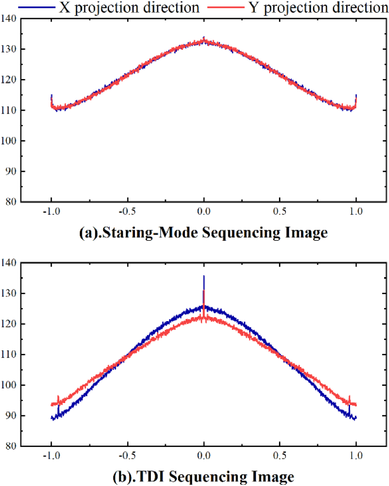
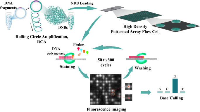
In high-throughput gene sequencing, the quality of sequencing images is critical for the accuracy of subsequent base calling. However, during practical sequencing processes, the time delay integration (TDI) camera's push-scan imaging often leads to significant degradation of image quality along the push-scan direction. Addressing the current limitations in TDI image restoration research for gene sequencing, this study establishes an imaging spectrum model of sequencing images based on MGI's ultra-high-throughput sequencer. We systematically analyze the causes and intrinsic mechanisms of image quality degradation, with a focus on elucidating the specific impacts of TDI push-scanning on image quality. To enhance TDI sequencing image quality, we compare the differences in power spectral projection curves between stare-mode imaging and TDI push-scan imaging and propose a power spectrum curve compensation (PSCC)-based quality optimization method alongside a novel evaluation framework for sequencing image quality. Experimental results demonstrate that compared to original H-channel images from cycle 1 to 50, the energy concentration (1/σ) of the optimized images increases by 9.13% in the TDI direction and 4.64% in the direction perpendicular to TDI. Signal-to-noise ratio (SNR) increases by 6.90% for base A and 4.99% for base C, while base calling accuracy (Q30) improves by 1.67%.
期刊介绍:
The journal''s scope encompasses fundamental research, technology development, biomedical studies and clinical applications. BOEx focuses on the leading edge topics in the field, including:
Tissue optics and spectroscopy
Novel microscopies
Optical coherence tomography
Diffuse and fluorescence tomography
Photoacoustic and multimodal imaging
Molecular imaging and therapies
Nanophotonic biosensing
Optical biophysics/photobiology
Microfluidic optical devices
Vision research.

 求助内容:
求助内容: 应助结果提醒方式:


