{"title":"Cone beam computed tomography analysis for immediate implant placement in the anterior maxilla based on radial root position: An observational study.","authors":"Jignasha Suthar, Somil Mathur, Snehal Upadhyay, Thanmai Taduri","doi":"10.4103/jips.jips_33_25","DOIUrl":null,"url":null,"abstract":"<p><strong>Aim: </strong>To validate the CBCT classification for immediate implant placement (IIP) given by Howard Gluckman in the local population of Gujarat, India, and additionally evaluate the available bone beyond the tooth apex for IIP in the direction of proposed osteotomy.</p><p><strong>Setting and design: </strong>Cross-sectional Observational study.</p><p><strong>Materials and methods: </strong>A total of 103 cone beam computed tomography (CBCT) scans involving the six maxillary anterior teeth were scrutinized in the radial plane. Each CBCT was divided into six slices (n = 618), which were classified according to Gluckman's classification, followed by making the osteotomy lines. Six measurements (L, W1, W2, W3, W4, and W5) were made from root to nasal floor. Bone length (L) was measured in the direction of proposed osteotomy, whereas the bone width was measured at five different points along the proposed osteotomy.</p><p><strong>Statistical analysis used: </strong>Chi-square p value, One-way ANOVA and Post hoc Tukey test.</p><p><strong>Results: </strong>As per Gluckman's classification, it was found that class I showed the highest bone width with the lowest bone length, whereas Class V showed the lowest bone width. The highest bone length was observed in Class IV. The prevalence of different radial root position (RRP) starting from class I to class V was 1%, 75%, 15%, 16%, and 3%, respectively.</p><p><strong>Conclusion: </strong>A distinct correlation was found between the anterior root position and the available bone between the root tip and the nasal floor as per Gluckman's classification.</p><p><strong>Clinical significance: </strong>This study helps in the radiographic evaluation of available bone around the roots of maxillary anterior teeth, which is a critical determining factor for treatment planning in IIP cases. A deep knowledge of RRP, bone morphology, and available alveolar bone beyond the apex provides useful perception to the clinician to plan surgical and grafting procedures to achieve primary stability. This will also help the clinicians to visualize the final prosthetic outcome with respect to the position of access hole.</p>","PeriodicalId":22669,"journal":{"name":"The Journal of Indian Prosthodontic Society","volume":"25 3","pages":"244-250"},"PeriodicalIF":1.0000,"publicationDate":"2025-07-01","publicationTypes":"Journal Article","fieldsOfStudy":null,"isOpenAccess":false,"openAccessPdf":"https://www.ncbi.nlm.nih.gov/pmc/articles/PMC12370110/pdf/","citationCount":"0","resultStr":null,"platform":"Semanticscholar","paperid":null,"PeriodicalName":"The Journal of Indian Prosthodontic Society","FirstCategoryId":"1085","ListUrlMain":"https://doi.org/10.4103/jips.jips_33_25","RegionNum":0,"RegionCategory":null,"ArticlePicture":[],"TitleCN":null,"AbstractTextCN":null,"PMCID":null,"EPubDate":"2025/7/16 0:00:00","PubModel":"Epub","JCR":"Q3","JCRName":"DENTISTRY, ORAL SURGERY & MEDICINE","Score":null,"Total":0}
引用次数: 0
Abstract
Aim: To validate the CBCT classification for immediate implant placement (IIP) given by Howard Gluckman in the local population of Gujarat, India, and additionally evaluate the available bone beyond the tooth apex for IIP in the direction of proposed osteotomy.
Setting and design: Cross-sectional Observational study.
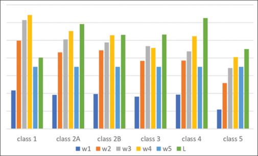
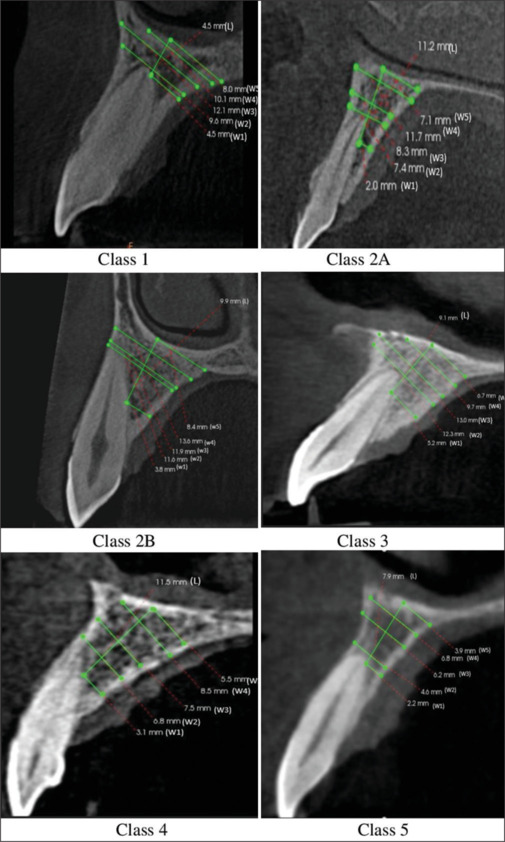
Materials and methods: A total of 103 cone beam computed tomography (CBCT) scans involving the six maxillary anterior teeth were scrutinized in the radial plane. Each CBCT was divided into six slices (n = 618), which were classified according to Gluckman's classification, followed by making the osteotomy lines. Six measurements (L, W1, W2, W3, W4, and W5) were made from root to nasal floor. Bone length (L) was measured in the direction of proposed osteotomy, whereas the bone width was measured at five different points along the proposed osteotomy.
Statistical analysis used: Chi-square p value, One-way ANOVA and Post hoc Tukey test.
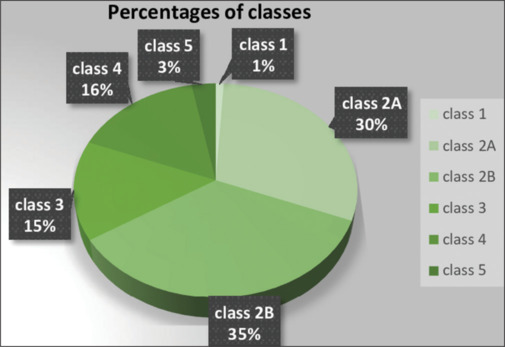
Results: As per Gluckman's classification, it was found that class I showed the highest bone width with the lowest bone length, whereas Class V showed the lowest bone width. The highest bone length was observed in Class IV. The prevalence of different radial root position (RRP) starting from class I to class V was 1%, 75%, 15%, 16%, and 3%, respectively.
Conclusion: A distinct correlation was found between the anterior root position and the available bone between the root tip and the nasal floor as per Gluckman's classification.
Clinical significance: This study helps in the radiographic evaluation of available bone around the roots of maxillary anterior teeth, which is a critical determining factor for treatment planning in IIP cases. A deep knowledge of RRP, bone morphology, and available alveolar bone beyond the apex provides useful perception to the clinician to plan surgical and grafting procedures to achieve primary stability. This will also help the clinicians to visualize the final prosthetic outcome with respect to the position of access hole.

 求助内容:
求助内容: 应助结果提醒方式:


