Tomas Vikner, Anders Garpebring, Cecilia Björnfot, Jan Malm, Anders Eklund, Anders Wåhlin
{"title":"MRI contrast accumulation in features of cerebral small vessel disease: blood-brain barrier dysfunction or elevated vascular density?","authors":"Tomas Vikner, Anders Garpebring, Cecilia Björnfot, Jan Malm, Anders Eklund, Anders Wåhlin","doi":"10.1186/s12987-025-00675-4","DOIUrl":null,"url":null,"abstract":"<p><strong>Background: </strong>White matter lesions (WML) and dilated perivascular spaces (PVS) are features of small vessel disease (SVD), commonly observed in aging and dementia, with unknown pathophysiology. Human studies have documented contrast accumulation within and in proximity of SVD-lesions. However, whether such observations mainly reflect excessive blood-brain barrier (BBB) leakage, or altered microvascular density in the investigated regions, remains unclear.</p><p><strong>Methods: </strong>To evaluate the roles of BBB leakage and vascular density in aging and SVD, dynamic contrast enhanced (DCE) MRI was used to estimate the permeability-surface area product (PS) and fractional plasma volume ([Formula: see text]) in normal-appearing brain tissue and in proximity of and within WML and PVS in a population-based cohort (N = 56; 34/22 m/f; age 64 to 84 years). Analysis of variance (ANOVA) was used to assess regional differences in PS and [Formula: see text] and analysis of covariance (ANCOVA) was used to assess regional differences in PS with [Formula: see text] and vascular risk as covariates.</p><p><strong>Results: </strong>Pronounced increases in PS and [Formula: see text] were observed from normal-appearing white matter (NAWM) to WML peripheries to WMLs. Similar PS and [Formula: see text]increases were observed from basal ganglia (BG) to BG-PVS. Further, PS in NAWM and white matter (WM) PVS were found to increase with cortex-to-ventricular depth. However, ANCOVA models with [Formula: see text] as a covariate showed that variance in PS was mainly explained by v<sub>p</sub> (η<sup>2</sup>=0.17 to η<sup>2</sup>=0.35; all p < 10<sup>- 3</sup>), whereas the effect of region was only borderline-significant when comparing NAWM, WML peripheries and WML (p = 0.03) and non-significant for the other comparisons (p > 0.29).</p><p><strong>Conclusions: </strong>Our findings support the notion that contrast leakage across the BBB accumulates within and in proximity of SVD-related lesions. However, high contrast accumulation may mainly reflect high vascularization, and to a lesser degree than previously recognized BBB dysfunction.</p>","PeriodicalId":12321,"journal":{"name":"Fluids and Barriers of the CNS","volume":"22 1","pages":"74"},"PeriodicalIF":6.2000,"publicationDate":"2025-07-16","publicationTypes":"Journal Article","fieldsOfStudy":null,"isOpenAccess":false,"openAccessPdf":"https://www.ncbi.nlm.nih.gov/pmc/articles/PMC12265124/pdf/","citationCount":"0","resultStr":null,"platform":"Semanticscholar","paperid":null,"PeriodicalName":"Fluids and Barriers of the CNS","FirstCategoryId":"3","ListUrlMain":"https://doi.org/10.1186/s12987-025-00675-4","RegionNum":1,"RegionCategory":"医学","ArticlePicture":[],"TitleCN":null,"AbstractTextCN":null,"PMCID":null,"EPubDate":"","PubModel":"","JCR":"Q1","JCRName":"NEUROSCIENCES","Score":null,"Total":0}
引用次数: 0
Abstract
Background: White matter lesions (WML) and dilated perivascular spaces (PVS) are features of small vessel disease (SVD), commonly observed in aging and dementia, with unknown pathophysiology. Human studies have documented contrast accumulation within and in proximity of SVD-lesions. However, whether such observations mainly reflect excessive blood-brain barrier (BBB) leakage, or altered microvascular density in the investigated regions, remains unclear.
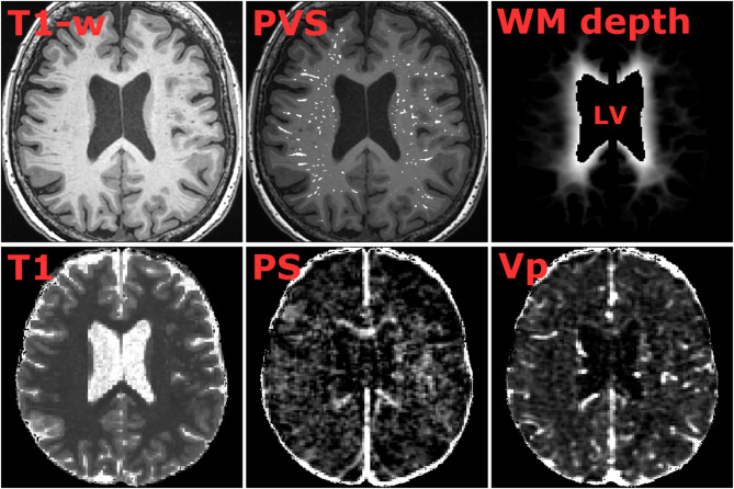
Methods: To evaluate the roles of BBB leakage and vascular density in aging and SVD, dynamic contrast enhanced (DCE) MRI was used to estimate the permeability-surface area product (PS) and fractional plasma volume ([Formula: see text]) in normal-appearing brain tissue and in proximity of and within WML and PVS in a population-based cohort (N = 56; 34/22 m/f; age 64 to 84 years). Analysis of variance (ANOVA) was used to assess regional differences in PS and [Formula: see text] and analysis of covariance (ANCOVA) was used to assess regional differences in PS with [Formula: see text] and vascular risk as covariates.
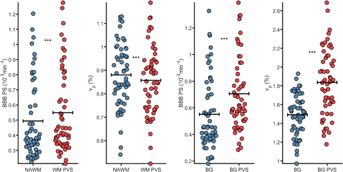
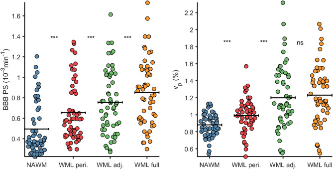
Results: Pronounced increases in PS and [Formula: see text] were observed from normal-appearing white matter (NAWM) to WML peripheries to WMLs. Similar PS and [Formula: see text]increases were observed from basal ganglia (BG) to BG-PVS. Further, PS in NAWM and white matter (WM) PVS were found to increase with cortex-to-ventricular depth. However, ANCOVA models with [Formula: see text] as a covariate showed that variance in PS was mainly explained by vp (η2=0.17 to η2=0.35; all p < 10- 3), whereas the effect of region was only borderline-significant when comparing NAWM, WML peripheries and WML (p = 0.03) and non-significant for the other comparisons (p > 0.29).
Conclusions: Our findings support the notion that contrast leakage across the BBB accumulates within and in proximity of SVD-related lesions. However, high contrast accumulation may mainly reflect high vascularization, and to a lesser degree than previously recognized BBB dysfunction.
期刊介绍:
"Fluids and Barriers of the CNS" is a scholarly open access journal that specializes in the intricate world of the central nervous system's fluids and barriers, which are pivotal for the health and well-being of the human body. This journal is a peer-reviewed platform that welcomes research manuscripts exploring the full spectrum of CNS fluids and barriers, with a particular focus on their roles in both health and disease.
At the heart of this journal's interest is the cerebrospinal fluid (CSF), a vital fluid that circulates within the brain and spinal cord, playing a multifaceted role in the normal functioning of the brain and in various neurological conditions. The journal delves into the composition, circulation, and absorption of CSF, as well as its relationship with the parenchymal interstitial fluid and the neurovascular unit at the blood-brain barrier (BBB).
 求助内容:
求助内容: 应助结果提醒方式:


