Megha R Kotecha, Varsha Manade, Jhimli Ta, Surbhi Chodvadiya
{"title":"Magnetic Extraction of an Intraretinal Foreign Body: A Case Report.","authors":"Megha R Kotecha, Varsha Manade, Jhimli Ta, Surbhi Chodvadiya","doi":"10.1159/000546787","DOIUrl":null,"url":null,"abstract":"<p><strong>Introduction: </strong>Inert intraocular foreign body (IOFB) removal depends on the location, type of injury, composition, and size of IOFB and possible serious complications of intraocular surgery. Early management is crucial for better prognosis.</p><p><strong>Case presentation: </strong>A 28-year-old male presented to the outpatient department after an alleged workplace accident. Initial assessment revealed significant diminution of vision, and on anterior segment examination with slit lamp, conjunctival congestion with no obvious entry point and no obvious scleral tear noted. Fundus examination by indirect ophthalmoscopy revealed vitreous hemorrhage, but the foreign body could not be localized due to extensive hazy media. Radiography of the orbit revealed an IOFB. The patient was managed surgically, and the intraretinal foreign body was removed using an intraocular magnet and intraocular forceps. The decision to remove the inert metal was considered because the patient had significant vision loss with vitreous hemorrhage.</p><p><strong>Conclusion: </strong>Management of an intraretinal metallic foreign body using an intraocular magnet is a viable and effective approach. It allows precise removal with minimal retinal trauma, thus preserving visual function.</p>","PeriodicalId":9635,"journal":{"name":"Case Reports in Ophthalmology","volume":"16 1","pages":"515-520"},"PeriodicalIF":0.6000,"publicationDate":"2025-06-14","publicationTypes":"Journal Article","fieldsOfStudy":null,"isOpenAccess":false,"openAccessPdf":"https://www.ncbi.nlm.nih.gov/pmc/articles/PMC12266699/pdf/","citationCount":"0","resultStr":null,"platform":"Semanticscholar","paperid":null,"PeriodicalName":"Case Reports in Ophthalmology","FirstCategoryId":"1085","ListUrlMain":"https://doi.org/10.1159/000546787","RegionNum":0,"RegionCategory":null,"ArticlePicture":[],"TitleCN":null,"AbstractTextCN":null,"PMCID":null,"EPubDate":"2025/1/1 0:00:00","PubModel":"eCollection","JCR":"Q4","JCRName":"OPHTHALMOLOGY","Score":null,"Total":0}
引用次数: 0
Abstract
Introduction: Inert intraocular foreign body (IOFB) removal depends on the location, type of injury, composition, and size of IOFB and possible serious complications of intraocular surgery. Early management is crucial for better prognosis.
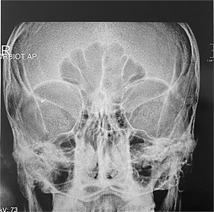
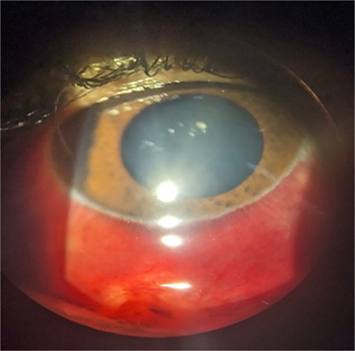
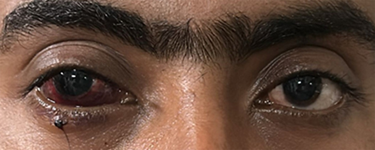
Case presentation: A 28-year-old male presented to the outpatient department after an alleged workplace accident. Initial assessment revealed significant diminution of vision, and on anterior segment examination with slit lamp, conjunctival congestion with no obvious entry point and no obvious scleral tear noted. Fundus examination by indirect ophthalmoscopy revealed vitreous hemorrhage, but the foreign body could not be localized due to extensive hazy media. Radiography of the orbit revealed an IOFB. The patient was managed surgically, and the intraretinal foreign body was removed using an intraocular magnet and intraocular forceps. The decision to remove the inert metal was considered because the patient had significant vision loss with vitreous hemorrhage.
Conclusion: Management of an intraretinal metallic foreign body using an intraocular magnet is a viable and effective approach. It allows precise removal with minimal retinal trauma, thus preserving visual function.
期刊介绍:
This peer-reviewed online-only journal publishes original case reports covering the entire spectrum of ophthalmology, including prevention, diagnosis, treatment, toxicities of therapy, supportive care, quality-of-life, and survivorship issues. The submission of negative results is strongly encouraged. The journal will also accept case reports dealing with the use of novel technologies, both in the arena of diagnosis and treatment. Supplementary material is welcomed. The intent of the journal is to provide clinicians and researchers with a tool to disseminate their personal experiences to a wider public as well as to review interesting cases encountered by colleagues all over the world. Universally used terms can be searched across the entire growing collection of case reports, further facilitating the retrieval of specific information. Following the open access principle, the entire contents can be retrieved at no charge, guaranteeing easy access to this valuable source of anecdotal information at all times.

 求助内容:
求助内容: 应助结果提醒方式:


