{"title":"Characterising corneal changes in aniridia-related keratopathy using in vivo confocal microscopy and a self-supervised AI model.","authors":"Abigail Eve Kaye, Yalin Zheng, Sajjad Ahmad","doi":"10.1136/bmjophth-2025-002300","DOIUrl":null,"url":null,"abstract":"<p><strong>Purpose: </strong>To investigate whether corneal changes observed via in vivo confocal microscopy (IVCM) in patients with aniridia-related keratopathy (ARK) reflect clinical severity.</p><p><strong>Methods: </strong>A cross-sectional, observational study. Patients with congenital aniridia and healthy controls were included. IVCM of the epithelium, anterior stroma and posterior stroma were collected, manually annotated and analysed using the pretrained DINOv2 model as a feature extractor. High-dimensional embeddings were visualised using t-distributed stochastic neighbour embedding (t-SNE) to assess layer-specific clustering. Structural deviations from normal controls were quantified using centroid and Euclidean distance metrics. The cumulative structural changes across corneal layers were then correlated with Ocular Surface Score (OSS), a clinical grading scale for ARK severity.</p><p><strong>Results: </strong>20 patients with congenital aniridia and six healthy controls were included. t-SNE analysis revealed distinct clusters for normal corneal layers; whereas, ARK samples displayed overlapping clusters, suggestive of blurred structural boundaries. Notably, significant clustering patterns were observed in the anterior stroma, even in cases with mild ARK, underscoring its potential as an early disease marker. Anterior stromal changes were significantly associated with OSS scores (p<0.05), while cumulative structural deviations across all layers demonstrated a stronger correlation with disease severity (p<0.01). The posterior stroma showed relative structural preservation, aligning closely with healthy controls.</p><p><strong>Conclusion: </strong>DINOv2 is a useful tool for identifying subtle structural changes in corneal layers affected by ARK. The corneal stromal features characterised using IVCM showed strong associations with clinical disease and may serve as structural biomarkers of clinical disease.</p>","PeriodicalId":9286,"journal":{"name":"BMJ Open Ophthalmology","volume":"10 1","pages":""},"PeriodicalIF":2.2000,"publicationDate":"2025-07-16","publicationTypes":"Journal Article","fieldsOfStudy":null,"isOpenAccess":false,"openAccessPdf":"https://www.ncbi.nlm.nih.gov/pmc/articles/PMC12273092/pdf/","citationCount":"0","resultStr":null,"platform":"Semanticscholar","paperid":null,"PeriodicalName":"BMJ Open Ophthalmology","FirstCategoryId":"1085","ListUrlMain":"https://doi.org/10.1136/bmjophth-2025-002300","RegionNum":0,"RegionCategory":null,"ArticlePicture":[],"TitleCN":null,"AbstractTextCN":null,"PMCID":null,"EPubDate":"","PubModel":"","JCR":"Q2","JCRName":"OPHTHALMOLOGY","Score":null,"Total":0}
引用次数: 0
Abstract
Purpose: To investigate whether corneal changes observed via in vivo confocal microscopy (IVCM) in patients with aniridia-related keratopathy (ARK) reflect clinical severity.
Methods: A cross-sectional, observational study. Patients with congenital aniridia and healthy controls were included. IVCM of the epithelium, anterior stroma and posterior stroma were collected, manually annotated and analysed using the pretrained DINOv2 model as a feature extractor. High-dimensional embeddings were visualised using t-distributed stochastic neighbour embedding (t-SNE) to assess layer-specific clustering. Structural deviations from normal controls were quantified using centroid and Euclidean distance metrics. The cumulative structural changes across corneal layers were then correlated with Ocular Surface Score (OSS), a clinical grading scale for ARK severity.
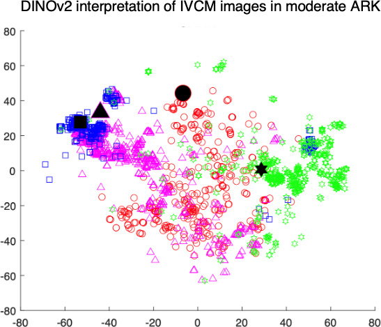
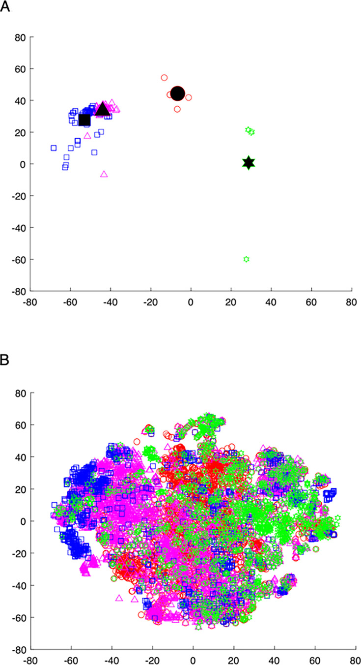
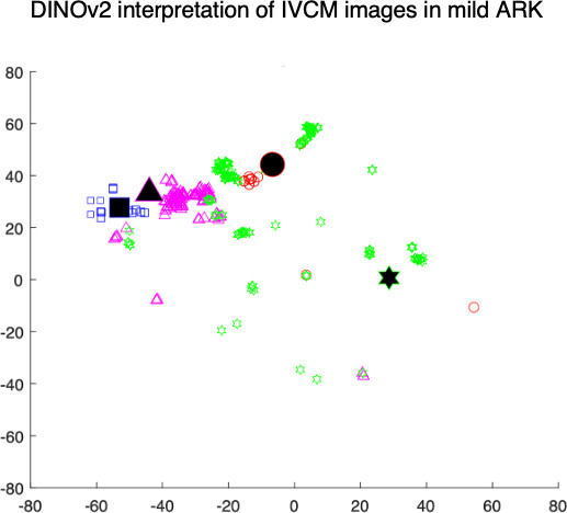
Results: 20 patients with congenital aniridia and six healthy controls were included. t-SNE analysis revealed distinct clusters for normal corneal layers; whereas, ARK samples displayed overlapping clusters, suggestive of blurred structural boundaries. Notably, significant clustering patterns were observed in the anterior stroma, even in cases with mild ARK, underscoring its potential as an early disease marker. Anterior stromal changes were significantly associated with OSS scores (p<0.05), while cumulative structural deviations across all layers demonstrated a stronger correlation with disease severity (p<0.01). The posterior stroma showed relative structural preservation, aligning closely with healthy controls.
Conclusion: DINOv2 is a useful tool for identifying subtle structural changes in corneal layers affected by ARK. The corneal stromal features characterised using IVCM showed strong associations with clinical disease and may serve as structural biomarkers of clinical disease.

 求助内容:
求助内容: 应助结果提醒方式:


