{"title":"Maxillary Cystic Ameloblastic Fibroma in an 8-Year-Old Girl: A Case Report Featuring a Rare Histological Variant.","authors":"Nasser Raqe Alqhtani","doi":"10.1155/crid/7645367","DOIUrl":null,"url":null,"abstract":"<p><p><b>Objective:</b> This report is aimed at outlining the unusual cystic variant of ameloblastic fibroma to facilitate its demarcation from other odontogenic lesions, including dentigerous cysts and cystic ameloblastomas. <b>Case Report:</b> An 8-year-old girl with no significant medical history presented to the oral surgery department with a painless swelling in the right maxillary region, first noticed 1 month ago, which gradually increased in size, accompanied by monocortical expansion of the buccal cortex. Cone beam computed tomography revealed a well-demarcated unilocular low-density lesion in the right posterior maxilla, measuring approximately 3 × 2 cm; the central bony lesion involved an unerupted first permanent molar. Conservative enucleation of the lesion was performed, along with the removal of the impacted tooth. Microscopic examination showed a benign mixed cystic odontogenic tumor, displaying odontogenic epithelial strands with stellate-shaped fibroblasts in a myxoid cell-rich stroma. The epithelial cells were rounded to cuboidal, with no mitotic activity or signs of malignancy. The overall histological image suggested a cystic ameloblastic fibroma. <b>Conclusion:</b> Clinically and radiographically, cystic ameloblastic fibroma may resemble a dentigerous cyst due to the involvement of an impacted tooth with the lesion. However, these two entities can be clearly histologically differentiated, as the distinctive odontogenic epithelial strands in a myxoid cell-rich stroma that are seen in cystic AF will be absent in a dentigerous cyst.</p>","PeriodicalId":46841,"journal":{"name":"Case Reports in Dentistry","volume":"2025 ","pages":"7645367"},"PeriodicalIF":0.9000,"publicationDate":"2025-07-08","publicationTypes":"Journal Article","fieldsOfStudy":null,"isOpenAccess":false,"openAccessPdf":"https://www.ncbi.nlm.nih.gov/pmc/articles/PMC12263257/pdf/","citationCount":"0","resultStr":null,"platform":"Semanticscholar","paperid":null,"PeriodicalName":"Case Reports in Dentistry","FirstCategoryId":"1085","ListUrlMain":"https://doi.org/10.1155/crid/7645367","RegionNum":0,"RegionCategory":null,"ArticlePicture":[],"TitleCN":null,"AbstractTextCN":null,"PMCID":null,"EPubDate":"2025/1/1 0:00:00","PubModel":"eCollection","JCR":"Q4","JCRName":"DENTISTRY, ORAL SURGERY & MEDICINE","Score":null,"Total":0}
引用次数: 0
Abstract
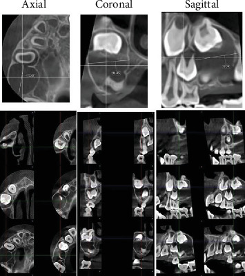
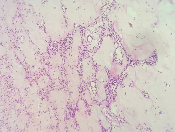
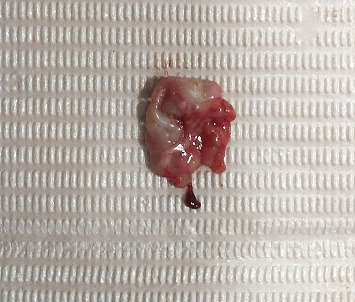
Objective: This report is aimed at outlining the unusual cystic variant of ameloblastic fibroma to facilitate its demarcation from other odontogenic lesions, including dentigerous cysts and cystic ameloblastomas. Case Report: An 8-year-old girl with no significant medical history presented to the oral surgery department with a painless swelling in the right maxillary region, first noticed 1 month ago, which gradually increased in size, accompanied by monocortical expansion of the buccal cortex. Cone beam computed tomography revealed a well-demarcated unilocular low-density lesion in the right posterior maxilla, measuring approximately 3 × 2 cm; the central bony lesion involved an unerupted first permanent molar. Conservative enucleation of the lesion was performed, along with the removal of the impacted tooth. Microscopic examination showed a benign mixed cystic odontogenic tumor, displaying odontogenic epithelial strands with stellate-shaped fibroblasts in a myxoid cell-rich stroma. The epithelial cells were rounded to cuboidal, with no mitotic activity or signs of malignancy. The overall histological image suggested a cystic ameloblastic fibroma. Conclusion: Clinically and radiographically, cystic ameloblastic fibroma may resemble a dentigerous cyst due to the involvement of an impacted tooth with the lesion. However, these two entities can be clearly histologically differentiated, as the distinctive odontogenic epithelial strands in a myxoid cell-rich stroma that are seen in cystic AF will be absent in a dentigerous cyst.
期刊介绍:
Case Reports in Dentistry is a peer-reviewed, Open Access journal that publishes case reports and case series in all areas of dentistry, including periodontal diseases, dental implants, oral pathology, as well as oral and maxillofacial surgery.

 求助内容:
求助内容: 应助结果提醒方式:


