{"title":"Methadone and the Kidney: Dissecting Gender Differences in Inflammation and Oxidative Stress Responses.","authors":"Katayoon Kosari, Shadan Saberi, Hamid Najafipour, Zoya Tahergorabi, Elham Jafari, Saeideh Jafarinejad Farsangi","doi":"10.34172/ahj.1625","DOIUrl":null,"url":null,"abstract":"<p><strong>Background: </strong>This study explored the gender-specific effects of methadone, a synthetic opioid receptor agonist commonly used in opioid addiction treatment, on renal tissue and function. We aimed to elucidate the underlying mechanisms involving inflammatory pathways and redox system activity.</p><p><strong>Methods: </strong>Forty-two Wistar rats (200-250 g) were allocated into six groups: three males and three females, each comprised of control, and methadone-treated 5 mg/kg and 20 mg/kg. Over eight weeks, animals received either saline or methadone syrup orally. Blood urea nitrogen (BUN) and serum creatinine (sCr) were measured in serum. The inflammatory cytokines and antioxidant enzyme activity were assessed in left kidneys, which were preserved at -80 °C, while histopathological analysis via H&E staining was done on the formalin-fixed right kidneys.</p><p><strong>Findings: </strong>Methadone administration resulted in renal tissue injury characterized by enhanced glomerular and interstitial inflammation. Notable increases in malondialdehyde (MDA), BUN, sCr, transforming growth factor beta (TGF-β), tumor necrosis factor alpha (TNF-α), and interleukin 17 (IL-17) were observed in methadone-treated groups, indicating impaired renal function associated with oxidative stress and inflammation, with male rats exhibiting more severe alterations. Conversely, methadone treatment elevated glutathione peroxidase (GPx), and catalase (Cat) activities, predominantly in females.</p><p><strong>Conclusion: </strong>Prolonged methadone therapy exerts a nephrotoxic effect through the activation of oxidative stress and inflammatory pathways, with male rats displaying greater renal pathology and dysfunction, potentially attributed to diminished antioxidant defenses.</p>","PeriodicalId":33943,"journal":{"name":"Addiction and Health","volume":"17 ","pages":"1625"},"PeriodicalIF":0.0000,"publicationDate":"2025-01-01","publicationTypes":"Journal Article","fieldsOfStudy":null,"isOpenAccess":false,"openAccessPdf":"https://www.ncbi.nlm.nih.gov/pmc/articles/PMC12260921/pdf/","citationCount":"0","resultStr":null,"platform":"Semanticscholar","paperid":null,"PeriodicalName":"Addiction and Health","FirstCategoryId":"1085","ListUrlMain":"https://doi.org/10.34172/ahj.1625","RegionNum":0,"RegionCategory":null,"ArticlePicture":[],"TitleCN":null,"AbstractTextCN":null,"PMCID":null,"EPubDate":"2025/5/14 0:00:00","PubModel":"Epub","JCR":"","JCRName":"","Score":null,"Total":0}
引用次数: 0
Abstract
Background: This study explored the gender-specific effects of methadone, a synthetic opioid receptor agonist commonly used in opioid addiction treatment, on renal tissue and function. We aimed to elucidate the underlying mechanisms involving inflammatory pathways and redox system activity.
Methods: Forty-two Wistar rats (200-250 g) were allocated into six groups: three males and three females, each comprised of control, and methadone-treated 5 mg/kg and 20 mg/kg. Over eight weeks, animals received either saline or methadone syrup orally. Blood urea nitrogen (BUN) and serum creatinine (sCr) were measured in serum. The inflammatory cytokines and antioxidant enzyme activity were assessed in left kidneys, which were preserved at -80 °C, while histopathological analysis via H&E staining was done on the formalin-fixed right kidneys.
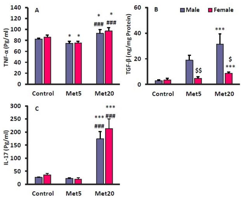
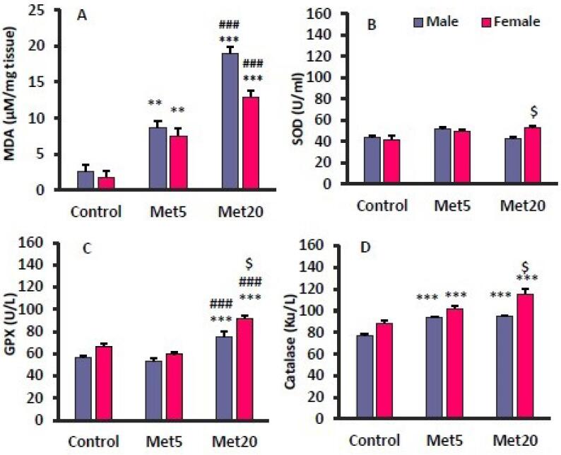
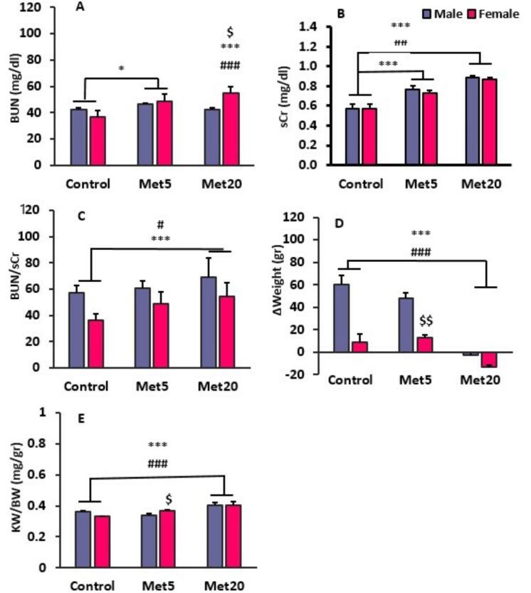
Findings: Methadone administration resulted in renal tissue injury characterized by enhanced glomerular and interstitial inflammation. Notable increases in malondialdehyde (MDA), BUN, sCr, transforming growth factor beta (TGF-β), tumor necrosis factor alpha (TNF-α), and interleukin 17 (IL-17) were observed in methadone-treated groups, indicating impaired renal function associated with oxidative stress and inflammation, with male rats exhibiting more severe alterations. Conversely, methadone treatment elevated glutathione peroxidase (GPx), and catalase (Cat) activities, predominantly in females.
Conclusion: Prolonged methadone therapy exerts a nephrotoxic effect through the activation of oxidative stress and inflammatory pathways, with male rats displaying greater renal pathology and dysfunction, potentially attributed to diminished antioxidant defenses.

 求助内容:
求助内容: 应助结果提醒方式:


